Figures & data
Figure 1. Flowchart of the experimental protocol.
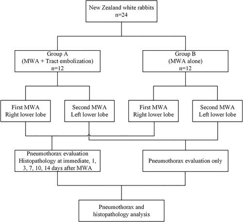
Figure 2. CT-guided lung microwave ablation and tract embolization in the right lower lobe. (A) A 15-G coaxial needle was introduced into the lung tissue ∼10 mm of the pleural surface under CT guidance. (B) Placement of the MW antenna. (C) Removal of the coaxial needle and sealing of the needle tract with gelatin sponge particle suspension (arrow). (D) CT performed 2 weeks after the procedure shows complete absorption of the gelatin sponge particles. The ablation zone is visible (arrowhead).
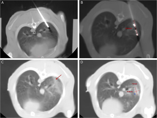
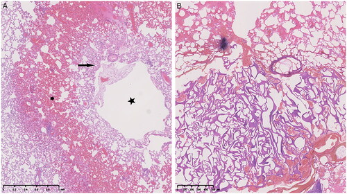
Table 1. Pneumothorax and aspiration rates in the two groups.
Figure 3. Rabbits were euthanized immediately after microwave ablation. (A) The histopathologic view (H and E, ×20) shows bilayer modifications around the needle tract: compressed and deformed alveolar walls in the inner layer (arrow), and congestion in the outer layer (arrowhead). Visualization of an open needle tract (star). (B) The slightly basophilic gelatin sponge particles fill the needle tract densely (H and E, ×40).