Figures & data
Table 1. Demographic characteristics of patients with vulvar lichen sclerosus (VLS).
Table 2. Comparison of treatment parameters and side effects between the two groups.
Figure 1. Side effects after FU treatment at 4.0 mm focal depth and the recovery of symptomatic treatment. (a) one patient’s vulvar lesion before FU treatment, (b) the same patient’s vulvar condition immediately after FU treatment, (c) the same patient’s ulcer 12 days after operation and (d) the same patient’s recovery after symptomatic treatment for 7 months after FU therapy.
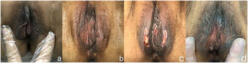
Figure 2. Side effects after FU treatment at 2.5 mm focal depth and the recovery of symptomatic treatment. (a) one patient’s vulvar lesion before FU treatment, (b) the same patient’s vulvar condition immediately after FU treatment, (c) the same patient’s ulcer 10 days after operation and (d) the same patient’s recovery after symptomatic treatment for 2 months after FU therapy.
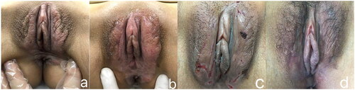
Table 3. Comparison of the efficacy of FU therapy between the two group.
Figure 3. Lesion size changed after FU treatment at 2.5 mm focal depth. (a) one patient’s vulvar lesion before FU treatment, (b) 7 days after FU treatment, (c) 3 months after FU treatment and (d) 6 months after FU treatment.
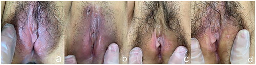
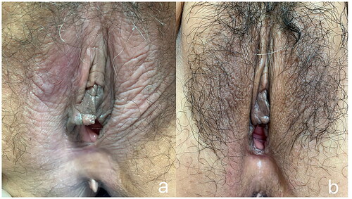
Figure 4. The lesion changed after FU treatment at 4.0 mm focal depth. (a) Clinical appearance before treatment. (b) The skin elasticity and pigmentation of the vulva were almost normal after 3 months of follow-up.