
Figures & data
Figure 1. Recurrent papillary thyroid cancer at left level 3 after thyroidectomy and RI therapy in a 35-year-old female. (a) Transverse ultrasound image showing a 0.7-cm-sized hypoechoic mass at right level 6 (arrows). (b) An internally cooled electrode with a 1.5-cm-sized active tip was inserted into the recurrent tumor (arrows). (c) One month after RFA, the US scans showed the treated tumor had decreased with 79% VRR (d). Six months after RFA, the treated tumor was not found on US. RI: radioactive iodine; RFA: radiofrequency ablation; US: ultrasound.
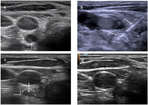
Table 1. Patient baseline characteristics.
Figure 2. Changes in the largest diameter before RFA and at each follow-up. RFA: radiofrequency ablation.
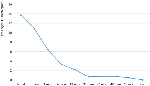
Figure 3. Changes in tumor volume before RFA and at each follow-up. RFA: radiofrequency ablation.
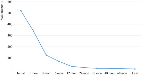
Figure 4. Changes in VRR before RFA and at each follow-up. RFA: radiofrequency ablation.
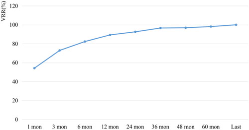
Table 2. Lesion characteristics.
Table 3. Long-term follow-up results of RFA.