
Figures & data
Figure 1. Pulmonary metastatic nodules on CT. CT images of the lung before chemotherapy, the largest pulmonary nodule is located in the left lung, with a diameter of 1.0 cm (A) and multiple nodules were seen (B). Shrinkage in the size of the pulmonary lesion pre-HIFU (C), the diameter of the lesion was 0.3 cm. Disappearance of the pulmonary nodules post-HIFU (D). CT, computed tomography; HIFU, high-intensity focused ultrasound.
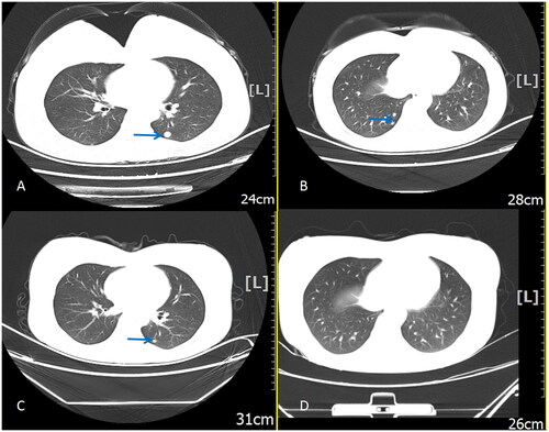
Figure 2. MRI of the uterine lesion pre-HIFU (A and B) and post-HIFU (C and D) treatment. Sagittal T2 image showing the uterine lesion of GTN located in the posterior wall and left horn of the uterus. The size was 3.6 cm. Flow void effect was seen (arrow pointing in Figure A) (A). Sagittal contrast-enhanced image showing the rich blood flow signal of the lesion (B). Sagittal contrast-enhanced imaging performed one day after the surgery showed no perfusion of uterine lesion (C). Sagittal contrast-enhanced image showing the shrinkage of the uterine lesion 22 days after HIFU treatment (size: 2.6 cm) (D). MRI, magnetic resonance imaging; HIFU, high-intensity focused ultrasound; GTN, gestational trophoblastic neoplasia.
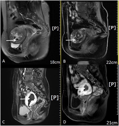
Figure 3. Ultrasound-guided HIFU treatment. Overall gray-scale change was seen during the surgery (A), while the vascular flow disappeared and the contrast-enhanced ultrasound showed no perfusion of uterine lesion immediately after the surgery (B). HIFU, high-intensity focused ultrasound.

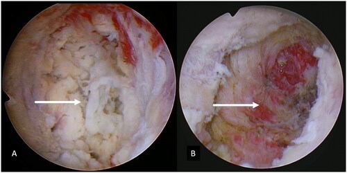
Figure 4. Hysteroscopic features after HIFU (A) and after hysteroscopic resection (B). White neoplastic tissue in the left horn of the uterus with no visual vascularity (A). The uterine lesion was completely resected under hysteroscopy (B). HIFU, high-intensity focused ultrasound; GTN, gestational trophoblastic neoplasia.
Availability of data and materials
The datasets used and/or analyzed during the current study are available from the corresponding author on reasonable request.