
Figures & data
Table 1. Baseline characteristics of the 32 patients with submucous leiomyomas who became pregnant after USgHIFU treatment.
Figure 1. A 33-year-old woman, with a history of miscarriage, achieved a successful full-term delivery via cesarean section after USgHIFU treatment for type 2 submucous leiomyoma. The maximum diameter was 4.2 cm, with an effective volume in the uterus cavity of 6.1 cm3 and a volume of submucous leiomyoma of 27.2 cm3. The contrast-enhanced ultrasound revealed no perfusion of uterine lesion immediately after the surgery (arrow pointing in Figure A). One year after USgHIFU treatment, both the effective volume in the uterine cavity and the volume of submucous leiomyoma shrank obviously (arrow pointing in Figure B in the sagittal plane and Figure C in the coronal plane). The maximum diameter was 2.4 cm, with an effective volume in the uterus cavity of 3.0 cm3 and a volume of submucous leiomyoma of 6.0 cm3. After 16 months, pregnancy was achieved. Gestational sac (blue arrow pointing in Figure D) was seen along with the reduced residual submucous leiomyoma, with a maximum diameter of 2.2 cm (white arrow pointing in Figure D). USgHIFU, ultrasound-guided high-intensity focused ultrasound.
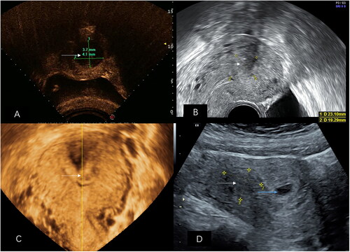
Table 2. Characteristics of submucous leiomyomas in 32 patients who became pregnant after USgHIFU treatment.
Table 3. HIFU treatment parameters for submucous leiomyomas in 32 patients who became pregnant.
Table 4. Pregnant characteristics of 32 pregnant women after USgHIFU treatment.
Table 5. Obstetric characteristics of 17 deliveries after USgHIFU treatment.
Data availability statement
The authors support data transparency.