
Figures & data
Figure 1. Classification of adenomyosis based on MRI. (A) Type I: the lesion invaded the endometrium layer, and the serosal layer (arrow) was intact; (B) Type II: the lesion invaded the outer uterine layer, the endometrium and the junctional zone (arrow) were intact; (C) Type III: lesions (arrows) confined to the myometrium with intact uterine structure; (D) Type IV: lesion (arrow) invaded the whole uterus.
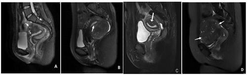
Table 1. SIR classification system for complications by the outcome.
Table 2. Baseline characteristics of patients with adenomyosis in four groups.
Table 3. Lesion characteristics and symptoms of the patients with adenomyosis in four groups [n(%)].
Table 4. The conditions required of individuals who completed the HIFU ablation in four groups (x ± s).
Table 5. Lesion size and menstruation volume before and after HIFU ablation in four groups (x ± s).
Table 6. The constituent ration of menorrhagia remission at 6 month after HIFU ablation in four groups [n(%)].
Table 7. The constituent ratio of dysmenorrhea relief at 6 month after HIFU ablation in four groups [n(%)].
Table 8. The constituent ratio of SIR scale after HIFU ablation in four groups [n(%)].
Table 9. The constituent ratio of after HIFU ablation in 227 patients.