Figures & data
Table 1. Mean CT number of normal liver tissue, standard deviation, and calculated upper and lower thresholds in Hounsfield units.
Figure 1. Semiautomatic segmentation steps. The volume of liver tissue without artifacts shown in red (A); The volume of interest encompassing ablation probe and artifacts within the liver in black (B); Voxels below the lower threshold in blue (C); Voxels above the upper threshold in brown (D); Union of voxels beyond the thresholds (E); Intersection of the volume of interest of the liver with the union of voxels beyond the thresholds resulting in artifact volume shown in yellow (F). Thresholds were calculated for each animal separately.
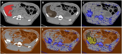
Figure 2. Examples of different artifact extents for qualitative evaluation of the liver parenchyma at probe tip; 1- marginal artifacts, 2-minor dark streaks < 2 cm, 3- distinct dark streaks > 2cm, 4- extensive artifacts hampering further puncture pathway planning.
Table 2. Mean values of artifact volumes, correction factors, and final artifact volume percentages of quantitative analysis.
Figure 3. Metal artifact shown in yellow in different ablation probes MWA (A,B), RFA (C,D) and CRYO (E,F) without iMAR (upper row) and with iMAR (bottom row).
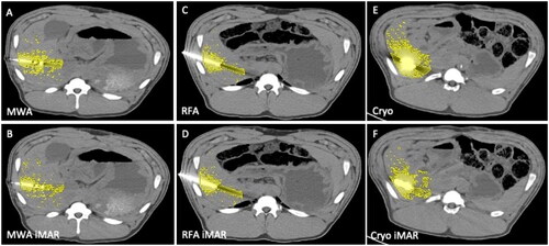
Table 3. Comparisons of metal artifact extent in different ablation probes are given in p-values with significant reduction of artifact volumes for the MWA probe (yellow boxes) and CRYO probe (green boxes). No significant artifact reduction was observed for RFA (blue boxes).
Figure 4. Box plots of noise (A) and contrast-to-noise (B) with median and upper and lower quartiles. No significant difference was seen between the ablation probes for noise and CNR. iMAR did not show significantly higher noise than non-iMAR images. No significant difference in CNR was found between probes, iterative reconstruction, or iMAR application.
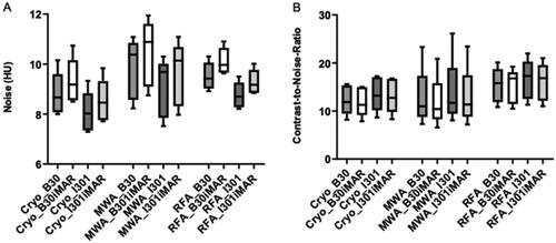
Table 4. Mean of subjective evaluation of overall metal artifacts and standard deviation to the 1st and 3rd percentiles in parentheses.
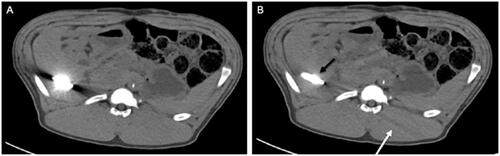
Figure 5. Images of a CRYO ablation probe without iMAR (A) and with iMAR (B). Blooming artifacts along the ablation probe (black arrow) and secondary artifacts in the image periphery detected in iMAR images (white arrow).