
Figures & data
Figure 1. Flowchart of patient selection.
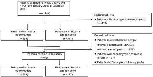
Figure 2. MRI features of internal and external adenomyosis. (A) Internal adenomyosis: a lesion with ill-defined margin located in the anterior wall of the uterus had developed in the thickened junctional zone (red arrow) and the myometrium outside the adenomyotic lesion was preserved. The thickness of the intact myometrium was 6 mm (white arrow). (B) External adenomyosis: a lesion with ill-defined margin (red arrow) located in the outer myometrium of the posterior wall of the uterus, the inner myometrium (the thickness was 8 mm, white arrow) between the adenomyotic lesion and the junctional zone was preserved, and the junctional zone was kept intact without aberrancy.
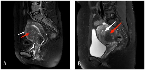
Figure 3. HIFU treatment for internal and external adenomyosis. (A) Pre-HIFU enhanced MRI showed an internal adenomyotic lesion located at the anterior wall of the uterus(arrow); (B) A contrast enhanced MR image obtained 1 day after HIFU showed the internal adenomyotic lesion was ablated(arrow). The NPV ratio was 89.5 %. (C) Pre-HIFU enhanced MRI showed an external adenomyotic lesion located at the posterior wall of the uterus(arrow); (D) A contrast enhanced MR image obtained 1 day after HIFU showed the external adenomyotic lesion was ablated without damaging to the serosa of the uterus(arrow). The NPV ratio was 44.1%.
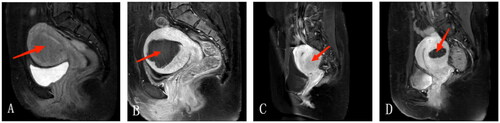
Table 1. Comparison of baseline characteristics of patients with internal and external adenomyosis.
Table 2. Comparison of HIFU treatment results between patients with internal and external adenomyosis.
Table 3. Comparison of baseline characteristics and HIFU treatment results between patients with internal and external adenomyosis in the posterior wall of the uterus.
Table 4. Comparison of adverse effects between patients with internal and external adenomyosis during HIFU treatment.
Table 5. Comparison of dysmenorrhea before and 18 month after HIFU between the two groups.
Table 6. Comparison of symptom improvement in patients with dysmenorrhea 18 month after HIFU between the two groups.
Table 7. Comparison of menorrhagia scores before and 18 month after HIFU between the two groups.
Table 8. Comparison of treatment efficacy in patients with menorrhagia 18 month after HIFU between the two groups.