
Figures & data
Table 1. Baseline characteristics of the patients.
Figure 1. Pre-HIFU, post-HIFU and follow-up MRI images obtained from a patient with type 1 submucosal fibroids. (A) The fibroids were located in the submucosa of the posterior wall of the uterus, with slightly high signal intensity on T2WI(Funaki III); (B) Moderate enhancement on T1WI; (C) 95% of the fibroids were ablated after HIFU, and there was no enhancement in the ablation area; Endometrium impairment grade 3(arrow); (D) Three months after HIFU, FVSR was 50%. Endometrium impairment grade 0(arrow).
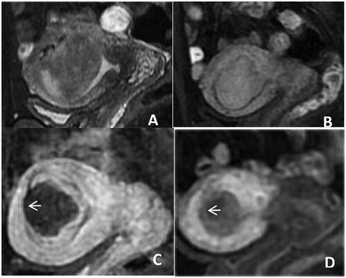
Figure 2. Pre-HIFU, post- HIFU and follow up HIFU MRI images obtained from a patient with type 2 submucosal fibroid. (A) The fibroids were located under the mucosa of the anterior wall of the uterus, with low signal intensity on T2WI(Funaki II); (B) Significant enhancement on T1WI; (C) 100% of the fibroids were ablated after HIFU, there is no enhancement in the mucosa of the fibroids; Endometrium impairmen grade 3(arrow); (D) 3 months after HIFU, FVSR was 90%, 100% enhancement of mucosa in fibroids. Endometrium impairmen grade 0(arrow).
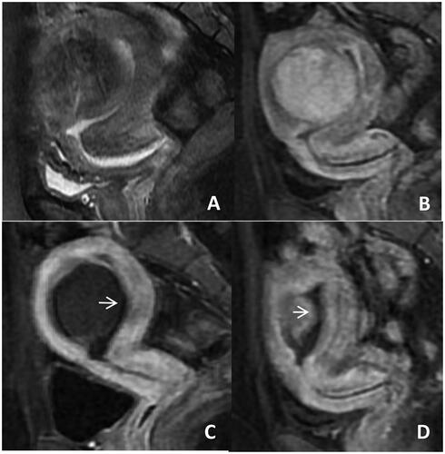
Figure 3. Pre-HIFU, post-HIFU and follow-up MRI images obtained from a patient with type 2–5 submucosal fibroids. (A) The fibroids were located under the mucosa of the anterior wall of the uterus, with low signal intensity on T2WI(Funaki I); (B) Slight to moderate enhancement after T1WI; (C) 100% of the fibroids were ablated after HIFU, there was no enhancement in the mucosa of the fibroids; Endometrium impairmen grade 3(arrow). (D) Three months after HIFU, FVSR was 50%. Excreted fibroid tissue was observed in the cervical canal. On the same day, the patient had abdominal pain and fibroid tissue was discharged through the vagina. Endometrium impairmen grade 3(arrow).
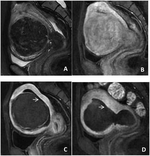
Table 2. Comparison of treatment efficacy.
Table 3. Analysis of submucosal fibroids type (FIGO) in FVSR, NPVR and endometrium impairment at three months after HIFU.