
Figures & data
Figure 1. The schematic diagram of percutaneous intrauterine instillation of chilled saline under the ultrasonic guidance for microwave ablation treating uterine fibroid adjacent to the endometrium. The submucous fibroid (red arrow); the 18 gauge puncture needle (blue arrows); the uterine cavity filled with chilled saline (green arrow).
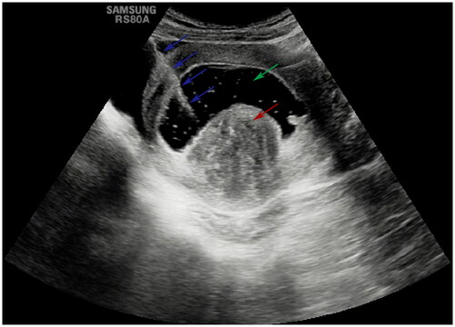
Table 1. The baseline characteristics of the study population.
Figure 2. The schematic diagram of endometrial impairment observed during postoperative hysteroscopy. (a) Normal endometrium: a 31-year-old patient with a type 2 fibroid; the endometrium was intact and the opening of the endometrial glands (blue arrows) could be observed. (b) Mild thermal damage: a 49-year-old patient with a type 3 fibroid; mild disorder, congestion and reddening of the endometrium (blue arrows) due to heat injury were observed. (c) Moderate thermal damage: a 39-year-old patient with a type 2 fibroid; a burnt necrosis with a size < 1 cm on the functional layer surface of the endometrium (blue arrow) was observed while the deep portion of the functional layer and the basal layer of the endometrium was intact (green arrow).
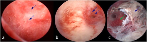
Figure 3. The schematic diagram of endometrial impairment grades indicated by red marks on postoperative magnetic resonance imaging. (a) Grade 0 (normal endometrium). (b) Grade 1 (pin-point, full-thickness discontinuity of involved endometrium). (c) Grade 2 (between grade 1 and 3). (d) Grade 3 (full-thickness discontinuity of involved endometrium over 1 cm in size).
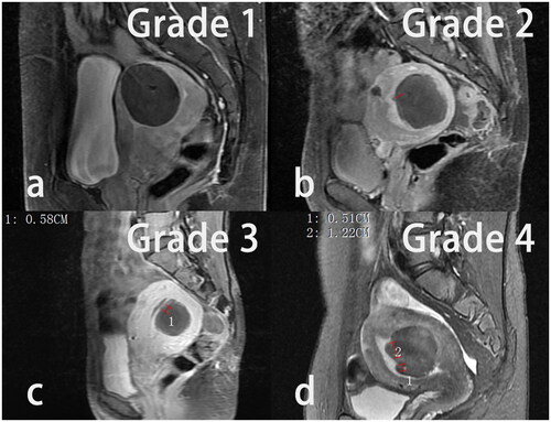
Table 2. The comparison of endometrial impairment between two groups.
Data availability statement
The data that support the findings of this study are available from the corresponding author upon reasonable request.