
Figures & data
Table 1. Scan parameters.
Table 2. Patient characteristics.
Table 3. Complications (SIR categories).
Figure 1. Pre-ablation imaging: a 60-year-old male patient with multifocal liver cancer recurred after TACE. Abnormal nodules were seen in the S4 and S5 segment. liver-specific contrast enhanced scan showed slightly low signal (a,b). the scanning equipment was Ge MR750W with the following parameters: FOV = 40 cm, phase FOV = 0.8, and layer thickness 5.0 mm. Matrix = 256 * 192, NEX = 1, bandwidth = 142.8, flip angle = 15.

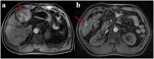
Figure 2. Intraoperative imaging: the microwave ablation needle showed low signal in all sequences and was inserted into the center of the tumor step by step (a,b). the scanning equipment was Ge MR750W with the following parameters: FOV = 40 cm, phase FOV = 0.8, and layer thickness 5.0 mm. Matrix = 256 * 192, NEX = 1, bandwidth = 142.8, flip angle = 15.
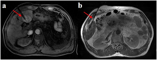
Figure 3. Post-ablation imaging: after the ablation was completed, the ablation lesion displayed a ‘target sign’ in the LAVA-Flex sequence (a,b). the scanning equipment was Ge MR750W with the following parameters: FOV = 40 cm, phase FOV = 0.8, and layer thickness 5.0 mm. Matrix = 256 * 192, NEX = 1, bandwidth = 142.8, flip angle = 15.