
Figures & data
Table 1. Patient characteristics.
Figure 1. Pretreatment MR-images. Histological samples are from the regions of the remaining leiomyoma tissue. CD: collagen% and CD31% show heterogeneous histology. Scale bar applies for all histological images.
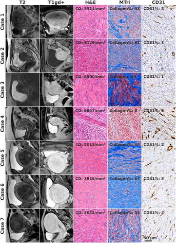
Table 2. Procedural outcomes.
Figure 2. (a) Samples from regions with dense scarred tissue. Organized collagen bundles mixed sparsely with basophilic nuclei are seen. Few blood vessels are present in case 7. Scale bar applies for all images. (b) Samples from regions with non-organized coagulative necrosis. Original tissue architecture is preserved, empty cell compartments and unorganized collagen bundles are seen. Hematoidin deposits and remnants of blood vessels are present. Scale bar applies for all images.
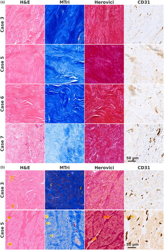
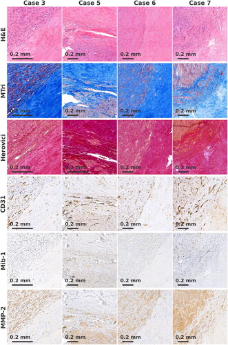
Figure 3. Boundary of remaining leiomyoma tissue and dense scarred tissue. No increase of Mib-1+, CD31+ or MMP-2+ cells is seen at the boundary. In Herovici, staining collagen appears mature.
Supplemental Material
Download Zip (25.8 MB)Data availability statement
Data are made available on reasonable request.