
Figures & data
Figure 1. Magnetic resonance image (MRI) obtained from a 26-year-old patient with diffuse uterine leiomyomatosis (DUL). (A) The pre-HIFU T2- weighted images showed that the uterus was pervaded with tumors of varied sizes in the myometrium. The size of the enlarged uterus, measuring 104 mm × 110mm × 103 mm, corresponded to that of 16 weeks of gestation. (B) Pre-HIFU contrast enhanced MRI revealed significant enhancement of leiomyomas. (C) Post-HIFU contrast enhanced MRI revealed non-perfused leiomyomas that distributed along the anterior wall. (D) Contrast enhanced MRI after second HIFU revealed non-perfused leiomyomas that distributed along both the anterior and posterior walls.
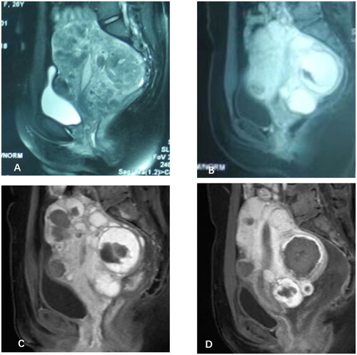
Figure 2. MRI obtained from a 32-year-old patient with DUL. (A and B) the T2WI and contrast-enhanced imaging revealed an enlarged uterus Filled with innumerable perfused fibroid nodules in each layer of the uterus before HIFU treatment. (C) MRI obtained one day after the first HIFU treatment show no enhancement of the leiomyomas at anterior uterine wall. (D) MRI obtained one day after the second HIFU treatment show no enhancement of leiomyomas at both the anterior and posterior walls of the uterus.

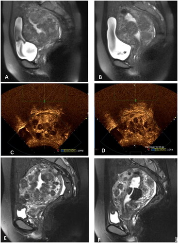
Figure 3. MRI obtained from a 27-year-old patient with DUL. (A&B) the pre-HIFU treatment T2-weighted imaging showed that the uterus,107mm × 85mm × 100 mm in size, was pervaded with tumors of varied sizes in the myometrium, which mainly was Isointense on T2WI. (C&D) the contrast-enhanced ultrasound showed non-perfusion in most of the leiomyomas immediately after HIFU treatment. (E&F)T2-weighted image revealed the uterus was 104 mm × 81mm × 89 mm in size, with a 17.6% shrinkage at 6 months compared to its initial volume prior to the treatment.
Data availability statement
The data that support the findings of this case report are available upon request from the corresponding author, LZ. The data are not publicly available because they contain information that can compromise the privacy of the research participants.