
Figures & data
Figure 1. Patient flowchart. Note: MWA = microwave ablation; SHPT = secondary hyperparathyroidism.

Figure 2. Ultrasound images of traditional hydrodissection and improved hydrodissection. (A) Traditional hydrodissection before ablation. A narrow anechoic isolating band (white thin arrows) was established outside the SHPT lesion (white thick arrow) by one-time injection. (B) During ablation, the isolating liquid (white thin arrows) was absorbed and diffused over time, there was no sufficient separating distance. (C) Improved hydrodissection before ablation. The pretracheal space (PTS), the retropharyngeal space (RS) and the anterior cervical space (ACS) were hydrodissected. The isolating fluid formed an anechoic isolating band (white thin arrows) and separated SHPT lesion (white thick arrow) as an ‘island’. (D) During ablation, the thickness of isolating band (white thin arrows) was maintained at least 5mm through continuous injection of NS. (E) Improved hydrodissection in the TGS (thin white arrows), which shows as a semicircular multilayer mixed-echoic area. (F) Improved hydrodissection in the CPS (thin white arrows). After injection of NS, the ‘onion skin’ sign emerged around SHPT lesion (white thick arrow), which showed a multiple layer and annular anechoic area. Note: THY, thyroid; ESO, esophagus; TRA, trachea; CCA, common carotid artery; IJV, internal jugular vein; CPS, circumferential periparathyroidal space; TGS, tracheoesophageal groove space = TGS.
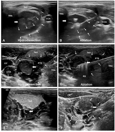
Figure 3. Ultrasound Image of hydrodissected TGS and CPS and the pathological images. (A) The TGS (white thin arrows) shows as a semicircular multilayer mixed-echoic area. (B) Pathological image shows corresponding fascia (white thin arrows) distribution in the tracheoesophageal groove area. (C) The CPS (white thin arrows) shows as an annular multilayer mixed-echoic area around SHPT lesion (white thick arrow). (D) The MASSON stains of resected SHPT lesion showed there were several layers of circular distributed collagen fibers (white thin arrows) around SHPT lesion (white thick arrow). Note: THY, thyroid; ESO, esophagus; TRA, trachea; CCA, common carotid artery; IJV, internal jugular vein; CPS, circumferential periparathyroidal space; TGS, tracheoesophageal groove space.
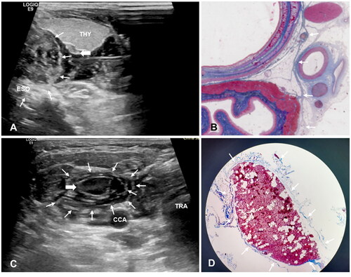
Table 1. Baseline characteristics.
Figure 4. The schematic diagram of different fascial spaces around parathyroid. The schematic diagram of hydrodissected PTS, RS, ACS, CS, CPS and TGS. Note: THY, thyroid; ESO, esophagus; TRA, trachea; CCA, common carotid artery; IJV, internal jugular vein; SHPT, secondary hyperparathyroidism; PTS, pretracheal space; RS, retropharyngeal space; ACS, anterior cervical space; CS, carotid space; CPS, circumferential periparathyroidal space; TGS, tracheoesophageal groove space.
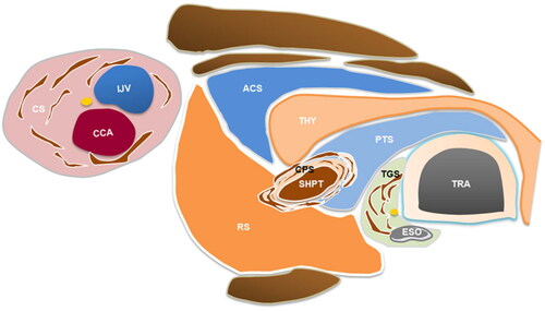
Figure 5. (A-F) Ultrasound images of SHPT lesions (White arrow) at different locations before and after improved hydrodissection. Pretracheal space hydrodissection (white arrowhead), retropharyngeal space hydrodissection (yellow arrowhead), anterior cervical space hydrodissection (black arrowhead), and carotid space hydrodissection (blue arrowhead) are shown as anechoic or mixed echoic bands on the images. Note: THY, thyroid; ESO, esophagus; TRA, trachea; CCA, common carotid artery; IJV, internal jugular vein; Sub-A, subclavian artery; SHPT, secondary hyperparathyroidism.
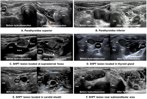
Table 2. The factors Influencing the development of hoarseness.
Data availability statement
Some or all datasets generated and/or analyzed during the current study are not publicly available but are available from the corresponding author upon reasonable request.