
Figures & data
Figure 1. Timeline. Timeline of first pig study (A). Timeline of second pig study using specialty diet during pig preparation (B).

Figure 2. Histotripsy Therapy System. Clinical histotripsy cart with 500 kHz transducer and coaxial US imaging probe mounted onto a micro-positioner (A) freehand imaging by a radiologist to identify acoustic window (B). UMC bowl with therapy transducer submerged via an adjustable arm (C).
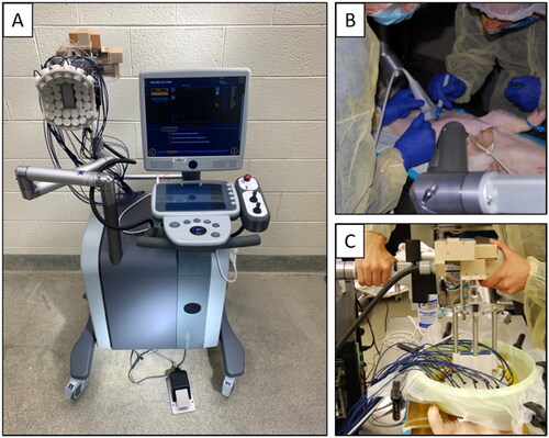
Table 1. Summary of all pig treatments. Note, regions of the pancreas visualized on US are in reference to the organ anatomy.
Figure 3. Histotripsy Treatment - Fasted Acute Pigs. Freehand US imaging of pancreas body indicated by red dashed lines in a fasted pig (A). US imaging with standoff showing gas obstruction prior to histotripsy treatment (B). Intestinal gas interference with treatment prevented ideal imaging quality and bubble formation during treatment leading to pre-focal intestinal bruising (C-D) more highly concentrated in the more gaseous large intestine, indicated by the green arrow, and small intestine, indicated by the blue arrows. Post-treatment H&E histopathology indicated that from a sample of the bruised colon, the muscularis was not damaged but there was a large amount of ablation and hemorrhage within the submucosa (E-F).
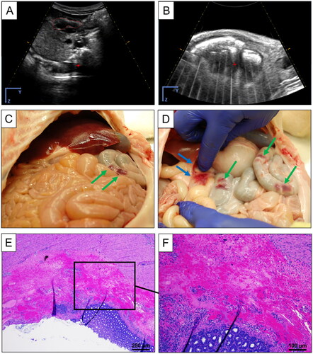
Figure 4. Histotripsy Treatment - Specialty Diet Acute Pigs. Freehand US imaging of pancreas before histotripsy indicated by dashed lines (A). US image with standoff prior to treatment and ROI identified by dashed lines (B). US image during treatment of bubble cloud generated in the pancreas (C) and post-treatment (D). Supplementary Video S1.
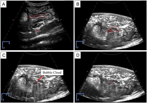
Figure 5. Histotripsy Treatment - Specialty Diet Chronic Pigs. US imaging of the pancreas before histotripsy indicated by dashed lines (A). US image with standoff prior to treatment and target identified by the dashed red circle (B). US image during treatment of bubble cloud generated in the pancreas (C) and after treatment with a clear hypoechoic region identifying the targeted ablated volume (D). Dashed red circle indicated the approximate region treated. Supplementary Video S2.
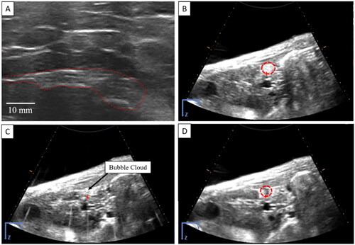
Figure 6. Necropsy - Specialty Diet Acute Pigs. Necropsy revealed no gross damage within the abdomen in situ (A). When excised in tota no damage was found on the exterior of the pancreas (B), nor other organs (liver, spleen, intestines) not shown, representative of pigs SD-1 and SD-2. With laxative, simethicone, and custard pretreatment protocol, there was significantly less bruising in prefocal bowel (C) indicated by the green arrow. Bruising around the treatment area was more concentrated than on the bowel but was still less intense than in the pilot group for pig SD-4. Histological staining of the pancreas revealed some loss of pancreatic tissue architecture at the periphery of lobules with the expansion of the interlobular tissue by flocculent debris, representative of pigs SD-3 and SD-4(D).
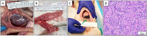
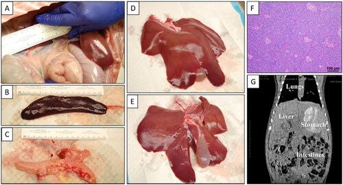
Figure 7. Necropsy - Specialty Diet Chronic Pigs. Necropsy revealed no gross damage within the abdomen in situ (A). When excised in tota no damage was found on the exterior of the spleen (B), pancreas (C), nor liver (D-E). 1-week post-treatment showed no signs of morbidity within the pancreatic tissue (F) and CT did not show any indication of ablation or off-target damage (G).
Supplemental Material
Download Zip (87.6 MB)Data availability statement
The data presented in this study are available on request from the corresponding authors.