
Figures & data
Table 1. Baseline characteristics of patients with low-risk PTC according to tumor location.
Figure 1. Kaplan–Meier analysis of local tumor progression-free survival in the isthmic and lobar groups before (A) and after (B) propensity-score matching.
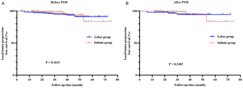
Table 2. Comparison of local tumor progression between the two groups.
Figure 2. Imaging findings in a 46-year-old Man with papillary thyroid cancer treated with radiofrequency ablation. The tumor located in the thyroid isthmus with a volume of 1.17 cm3 (arrow) is shown in the longitudinal (A) and transverse (B) planes on ultrasonography. The tumor volume decreased to 0.76 cm3 (arrow, C), 0.26 cm3 (arrow, D), and 0.17 cm3 (arrow, E), respectively, at 1, 3, and 9 months after radiofrequency ablation and disappeared at 18 months (arrow, F).
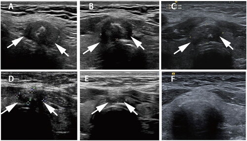
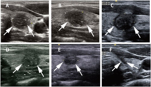
Figure 3. Imaging findings in a 30-year-old Woman with papillary thyroid cancer treated with radiofrequency ablation. The tumor located in the left lobe of the thyroid with a volume of 0.88 cm3 (arrow) is shown in the longitudinal (A) and transverse (B) planes on ultrasonography. The tumor volume decreased to 0.63 cm3 (arrow, C), 0.47 cm3 (arrow, D), and 0.33 cm3 (arrow, E), respectively, at 1, 3, and 6 months after radiofrequency ablation, and only the needle track was visible at 12 months (arrow, F).
Supplemental Material
Download ()Data availability statement
Some or all datasets generated during and/or analyzed during the current study are not publicly available but are available from the corresponding author upon reasonable request.