
Figures & data
Figure 1. Axial CT images of a 64-year-old man with stage IA NSCLC (adenocarcinoma) with IPF who underwent CT-guided synchronous percutaneous CNB and MWA. (a) CT prior to MWA treatment shows a 1.9 × 1.2 cm lesion in the right lower lobe (arrow); (b), (c) CT findings during MWA treatment, a 15 G coaxial introducer needle (arrow) was advanced to the proximal edge of the lesion, and the antenna was positioned centrally through the cannula into the lesion (arrow); (d) CT image immediately post-procedure showed ground-glass opacity around the tumor (arrow); (e) CT image 24 h post-procedure showed the expected thermal damage around the target lesion, without pneumothorax. (f) CT image 6 months after MWA treatment showed a reduced ablation area (arrow), cavitation changes in the primary focus, and surrounding fibrotic scarring with signs of inflammation. CT: computed tomography; NSCLC: non-small cell lung cancer; IPF: idiopathic pulmonary fibrosis; CNB: core-needle biopsy; MWA: microwave ablation.
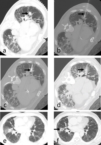
Figure 2. Axial CT images of a 76-year-old man with stage IA NSCLC (squamous carcinoma) with IPF who underwent CT-guided synchronous percutaneous CNB and MWA. (a) CT prior to MWA treatment showed a 1.4 × 0.9 cm subpleural lesion in the left upper lobe (arrow); (b) CT findings during MWA treatment, the antenna punctured the Central position of the lesion through honeycomb lesions (arrow); (c) CT image 24 h post-procedure showed ground-glass opacity around the tumor (arrow); (d) the 12-month CT image after MWA treatment revealed gradual shrinkage of the ablated lesion, and it became a fiber scar (arrow). CT: computed tomography; NSCLC: non-small cell lung cancer; CNB: core-needle biopsy; MWA: microwave ablation; IPF: idiopathic pulmonary fibrosis.
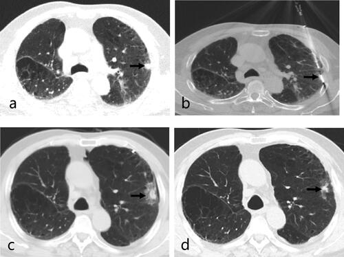
Table 1. Patients and tumor characteristics.
Table 2. Grade of complications during and following MWA.
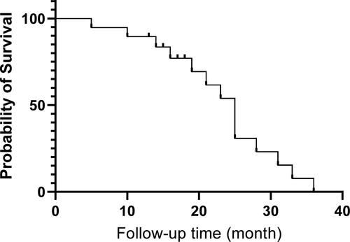
Figure 3. Kaplan-Meier survival curve for nineteen stage I non-small cell lung cancer (NSCLC) patients with idiopathic pulmonary fibrosis (IPF) that received microwave ablation (MWA) treatment.
Data availability statement
In addition to the raw data in the manuscript, the datasets used are available from the corresponding author on reasonable request.