
Figures & data
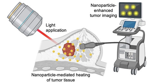
Scheme 1. Overview of theranostic NPs for dual cancer imaging and PTT. (A) Depiction of the process to use NPs for real-time imaging to guide PTT. (B) Summary of various NP formulations that have used pre-clinically as phototheranostics. Red spheres in the polymer, lipid and silica formulations indicate encapsulated light-responsive dyes. Created with BioRender.com.
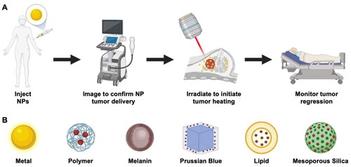
Scheme 2. Cell-derived membranes include distinct molecules that facilitate immune evasion, enable binding to non-homotypic cells or promote binding to homotypic cells. Together, these features enhance the tumor accumulation of MWNPs. Created with BioRender.com.
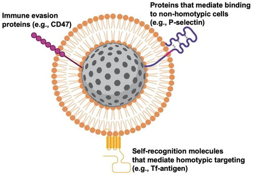
Table 1. List of common abbreviations found throughout this review.
Table 2. Comparison of different imaging modalities that have been utilized in combination with phototheranostic NPs.
Figure 1. Representative MWNP used for PTT, PA imaging and FL imaging. (A) Scheme of ICNP. (B) FL and PA images of MCF-7 breast cancer tumors in mice following IV injection with either ICG, INPs or ICNPs. (C) Thermal images of MCF-7 tumor-bearing mice exposed to an 808 nm laser for 5 min (1 W/cm2) after IV injection of PBS, ICG, INPs or ICNPs. (D) Tumor growth curves of different groups after treatments (n = 5). Reprinted (adapted) with permission from Chen et al. [Citation8,p.10049]. Copyright 2016 American Chemical Society.
![Figure 1. Representative MWNP used for PTT, PA imaging and FL imaging. (A) Scheme of ICNP. (B) FL and PA images of MCF-7 breast cancer tumors in mice following IV injection with either ICG, INPs or ICNPs. (C) Thermal images of MCF-7 tumor-bearing mice exposed to an 808 nm laser for 5 min (1 W/cm2) after IV injection of PBS, ICG, INPs or ICNPs. (D) Tumor growth curves of different groups after treatments (n = 5). Reprinted (adapted) with permission from Chen et al. [Citation8,p.10049]. Copyright 2016 American Chemical Society.](/cms/asset/6564b6a1-f7d5-46ed-aa22-060a2d4719ef/ihyt_a_2272066_f0001_c.jpg)
Figure 2. Representative example of membrane-wrapped semiconducting material for combined PTT and PA imaging. (A) Hybrid membrane-coated DOX-loaded hollow cupper sulfide NPs (DCuS@[RBC–B16] NPs) were explored for combination imaging/PTT/chemotherapy of melanoma. (B) CuS@[RBC–B16] NPs achieved a strong localized PA signal in tumors 4 h post IV injection. (C) Blood retention time of CuS NPs that were unwrapped or coated with RBC membranes, B16-F10 membranes or hybrid RBC–B16 membranes. (D) Biodistribution of unwrapped, RBC-wrapped, B16-wrapped or hybrid membrane-wrapped CuS NPs in tumor-bearing mice after IV injection. (E) Relative tumor volume of mice exposed to different treatments (1: NS, 2: CuS@[RBC–B16], 3: DOX, 4: NIR laser (1064 nm, 1 W/cm2), 5: DCuS@[RBC–B16], 6: CuS@[RBC–B16] with NIR laser, 7: DCuS@[RBC–B16] with NIR laser). Reprinted (adapted) with permission from Wang et al. [Citation25,p.5241]. Copyright 2018 American Chemical Society.
![Figure 2. Representative example of membrane-wrapped semiconducting material for combined PTT and PA imaging. (A) Hybrid membrane-coated DOX-loaded hollow cupper sulfide NPs (DCuS@[RBC–B16] NPs) were explored for combination imaging/PTT/chemotherapy of melanoma. (B) CuS@[RBC–B16] NPs achieved a strong localized PA signal in tumors 4 h post IV injection. (C) Blood retention time of CuS NPs that were unwrapped or coated with RBC membranes, B16-F10 membranes or hybrid RBC–B16 membranes. (D) Biodistribution of unwrapped, RBC-wrapped, B16-wrapped or hybrid membrane-wrapped CuS NPs in tumor-bearing mice after IV injection. (E) Relative tumor volume of mice exposed to different treatments (1: NS, 2: CuS@[RBC–B16], 3: DOX, 4: NIR laser (1064 nm, 1 W/cm2), 5: DCuS@[RBC–B16], 6: CuS@[RBC–B16] with NIR laser, 7: DCuS@[RBC–B16] with NIR laser). Reprinted (adapted) with permission from Wang et al. [Citation25,p.5241]. Copyright 2018 American Chemical Society.](/cms/asset/25378c30-f5df-45b5-80ec-deaae691dd12/ihyt_a_2272066_f0002_c.jpg)
Figure 3. Example of a MWNP used for dual FL imaging and PTT. (A) NIR-II FL images of mice bearing glioblastoma tumors following IV injection with either IR792, DINPs (unwrapped NPs loaded with IR792) or MDINPs (macrophage membrane-wrapped NPs loaded with IR792). (B) Representative bioluminescence images of tumors after treatment with different agents combined with 808 nm irradiation. (C) Survival rates of mice in different groups after PTT (n = 5). Reprinted (adapted) with permission from Lai et al. [Citation13,p.48]. Copyright 2019 American Chemical Society.
![Figure 3. Example of a MWNP used for dual FL imaging and PTT. (A) NIR-II FL images of mice bearing glioblastoma tumors following IV injection with either IR792, DINPs (unwrapped NPs loaded with IR792) or MDINPs (macrophage membrane-wrapped NPs loaded with IR792). (B) Representative bioluminescence images of tumors after treatment with different agents combined with 808 nm irradiation. (C) Survival rates of mice in different groups after PTT (n = 5). Reprinted (adapted) with permission from Lai et al. [Citation13,p.48]. Copyright 2019 American Chemical Society.](/cms/asset/949305ad-9985-4613-b80f-4e9141bb11fe/ihyt_a_2272066_f0003_c.jpg)
Table 3. Summary of studies that have developed MWNPs for image-guided PTT.
Data availability statement
Data sharing is not applicable to this article as no new data were created or analyzed in this study.