
Figures & data
Figure 1. Research flowchart.
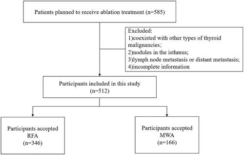
Figure 2. A case of RFA. (A) A 34-year-old female patient with a suspected lesion located in the right lobe of the thyroid, which was confirmed as PTMCs by FNAB; (B) After US-guided RFA, the ablated lesion was evaluated with CEUS;(C) In the 1st week follow up, the ablated lesion appeared as a well-defined heterogeneous echogenic area on the grayscale US; (D) On the final follow-up, the ablated area was almost completely absorbed.
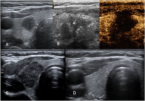
Figure 3. A case of MWA. (A) A 37-year-old female patient with a suspected lesion located in the right lobe of the thyroid, which was confirmed as PTMCs by FNAB; (B) After US-guided MWA, the ablated lesion was evaluated with CEUS; (C) In the first week follow up, the ablated lesion appeared as a well-defined heterogeneous echogenic area on the grayscale US with a central hypoechoic ablated needle channel; (D) On the final follow-up, the ablated area was almost completely absorbed.
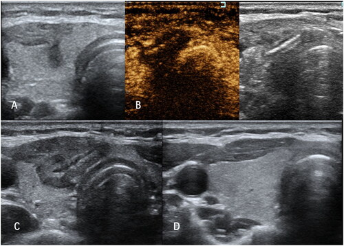
Table 1. Baseline characteristics of the patients between the two groups.
Table 2. Ultrasonographic characteristics of the nodules between the two groups.
Figure 4. The diagram displayed the VRR at each follow-up after RA or MWA. “**” indicates P < 0.01, “*” indicates P < 0.05.
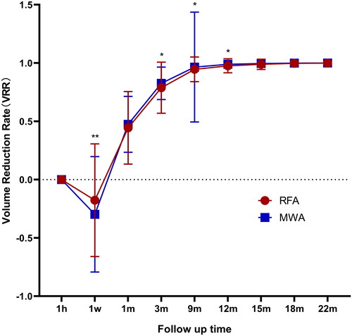
Table 3. Volumes of the ablated lesions between the two groups.
Table 4. Treatment parameters and complications in the two groups.
Data availability statement
There are ethical restrictions on sharing of de-identified data for this study. The ethics committee has not agreed to the public sharing of data as we do not have the participants’ permission to share their anonymous data