Figures & data
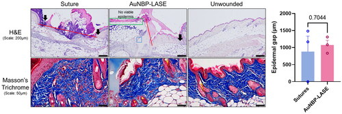
Figure 1. Spectroscopic characterization of gold nanobipyramids (AuNBPs). (a) A representative absorption spectrum of the AuNBP seed solution. (b) Normalized AuNBP absorption spectra as a function of seed volume used in a 4 mL reaction (with 8-HQL as a reducing agent). (c) Normalized absorption spectra of AuNBPs for ascorbic acid or 8-HQL as reducing agents. (d) TEM image of high-purity AuNBPs indicates a uniform size distribution. A representative image from n = 3 independent TEM experiments is shown. Additional TEM images are included in Supplementary Figure S1.
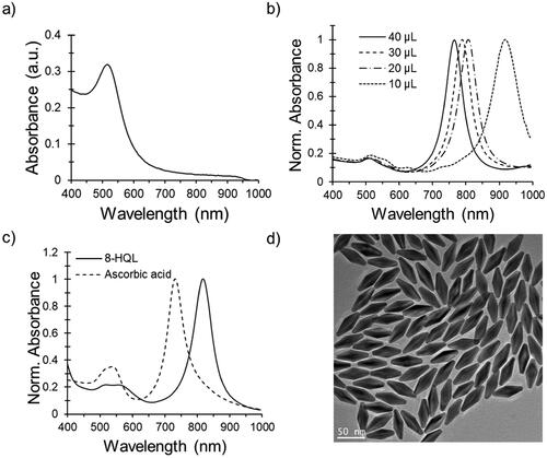
Figure 2. Characterization of AuNBP-LASE films. (a) Representative digital photograph of an AuNBP-LASE film prepared by solvent evaporation. (b) AuNBP absorption spectra normalized with maximum absorption in each spectrum as a function of hydration with saline (average of n = 3). (c) A representative absorption spectrum of dry AuNR-LASE. (d) photothermal (temperature) response of AuNBP-LASE and e) AuNR-LASE irradiated with an 808 nm laser at different power densities. The laser was irradiated (“on”) for 30 s and turned off (“off”) for 30 s for three consecutive cycles. Representative data from n = 3 independent experiments. (f) Dynamic mechanical analysis (DMA) of AuNBP-LASE (Average of n = 3).
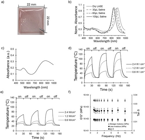
Figure 3. Skin sealing efficacy with AuNBP-LASE. (a) Digital photograph of an AuNBP-LASE used in laser skin sealing (left). Murine skin incisions sealed with sutures and AuNBP-LASE are shown on the right as indicated. (b) A cartoon showing vapometer and cutometer measurement sites in mice whose incisional wounds were laser-sealed with AuNBP-LASE. (c) Transepidermal water loss (TEWL) measurements of skin incisions sealed with sutures and AuNBP-LASE on day 2 post wounding d) Representative cutometer curves of the healing skin on day 2 post wounding using Mode 1 of the cutometer. (e) Comparison of dimensionless parameters obtained from cutometer measurements for unwounded skin, suture-approximated skin, and AuNBP-LASE-sealed skin. Descriptions of individual parameters are provided in . Representative images and data are shown from at least n = 3 independent experiments.
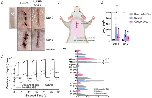
Table 1. Description of cutometer parameters.
Figure 4. Histological analyses of mice incisions closed with sutures and AuNBP-LASE on day 2 post wounding. Representatives of n = 3 independent images are shown here; other images are shown in Supplementary Figure S4. (Top) Hematoxylin and eosin (H&E) staining (red lines indicate the dermal gap, black arrows indicate the epidermal edge, blue box shows the area of glue integration with the tissue, and green box illustrates the effect of laser-induced hyperthermia on dermal collagen) and (bottom) Masson’s trichrome staining. (Right) Quantification of epidermal gap for incisions closed using sutures and AuNBP-LASE.