
Figures & data
Figure 1. Flowchart illustrating patient selection criteria.
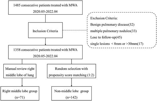
Table 1. Baseline characteristics of patients and pulmonary nodules compared between the RML and non-RML groups.
Table 2. Complications and post-ablation hospital stay compared between the RML and non-RML groups.
Figure 2. A female, 55-year-old patient with GGN-like lung cancer in the RML underwent MWA. (A) A mixed GGN located in the RML with a diameter of 12 mm. (B) An antenna was inserted into the tumor. (C) The lesion was covered by ground-glass opacity, and a small amount of pneumothorax was observed immediately post-ablation. (D) The lesion involuted at 1-month post-ablation. (E) The lesion gradually involuted at 12-months post-ablation. (F) The lesion involuted into a band at 36-months post-ablation.
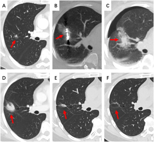
Figure 3. A male, 49-year-old patient with GGN-like lung cancer in the RML underwent MWA. (A) A mixed GGN located in the RML with a diameter of 11 mm. (B) An antenna was inserted into the tumor. (C) A small amount of pneumothorax occurred immediately after MWA. (D) The lesion was covered by ground-glass opacity, displaying a “fried egg” sign, and the pneumothorax volume did not increase. (E) The lesion gradually involuted at 6-months post-ablation. (F) The lesion involuted into a fibrous cord at 14-months post-ablation.
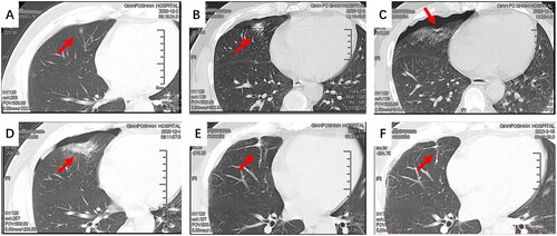
Figure 4. A female, 65-year-old patient with GGN-like lung cancer in the RML underwent MWA. (A) A mixed GGN located in the RML with a diameter of 17 mm. (B) An antenna was inserted into the tumor through the interlobar fissure. A large amount of pneumothorax occurred during the procedure, causing tumor displacement. (C) Continuous negative-pressure chest drainage was performed. (D) After negative-pressure aspiration, the procedure was continued. (E) The lesion was covered by ground-glass opacity, displaying a “fried egg” sign, and the amount of pneumothorax and subcutaneous emphysema did not increase at 72 h after MWA. (F) The lesion gradually involuted at 1 month after MWA. (G) The lesion gradually involuted at 10 months after MWA. (H) The lesion gradually involuted into a fibrous scar at 25 months after MWA.
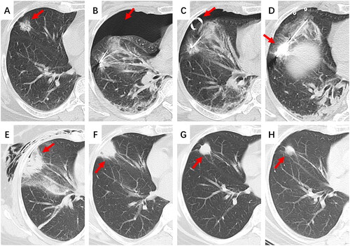
Table 3. Univariate and multivariate conditional logistic regression analysis of risk factors for pneumothorax.
Data availability statement
The data that support the findings of this study are available on request from the corresponding author, Xin Ye. The data are not publicly available due to their containing information that could compromise the privacy of research participants.