
Figures & data
Figure 1. Model geometry used to simulate ablation with a dMWA device in the vertebral body of a 45 kg pig.
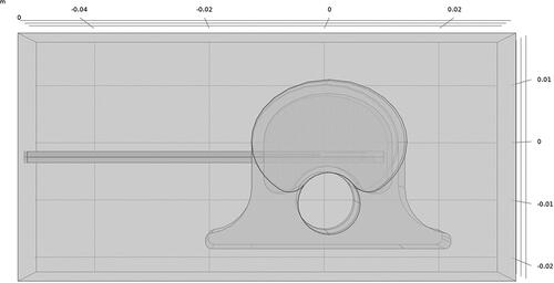
Table 1. Tissue biophysical properties employed for modeling 2.45 GHz microwave ablation in ex vivo porcine vertebral body [Citation15].
Figure 2. Ex vivo porcine model. Placement of temperature sensors (left) and dMWA applicator (right) within vertebral body.
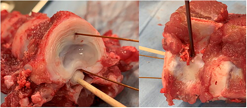
Figure 3. Image of vertebral body following ex vivo microwave ablation (H&E stained section). Optimal T1 placement distance (A); optimal T2 placement distance (B); extent of nuclear streaming/precipitation (necrosis) (C); applicator tract width (D). Ventral surface (V), vertebral foramen/dorsal aspect (VF), ablation direction (green arrow).
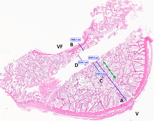
Figure 4. 80 W, 3.5 min ex vivo ablation simulation with initial tissue temperature of 20 °C.
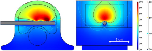
Table 2. Simulated ex vivo bone ablation results.
Figure 5. Experimentally measured T1 (Ch1) and T2 (Ch2) during 80 W, 3.5 min ablation (left) and during 120 W, 5 min ablation (right) in ex vivo porcine vertebral body.
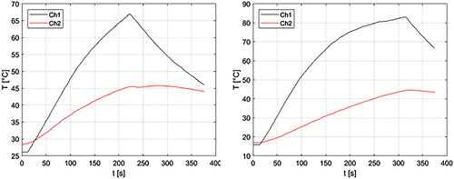
Table 3. Ex vivo vertebrae ablation experimental results (mean ± std).
Table 4. Ex vivo vertebra ablation experimental vs. simulated ΔT1 and ΔT2.
Figure 6. Transverse cut of vertebra, along the DMWA applicator pathway. Ablation conditions: 120 W, 5 min, room temperature initial temperature ex vivo ablation zone (A) Corresponding H&E stained section (B).
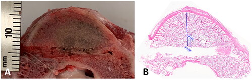
Figure 7. Sections of ablated and unablated vertebrate. Area immediately adjacent to DMWA applicator antenna heating showing severe bone marrow coagulation necrosis characterized by complete loss of cellular detail, as well as precipitation and clumping of cell remnants, coagulation of serum, erythrocytes, and marrow stroma (A). Area adjacent to (A) exhibiting less severe coagulative necrosis characterized by diffuse pallor and only partial loss of cellular details (individual cells and their nuclear detail are retained), as well as mild precipitation and clumping of cell remnants, coagulation of serum, erythrocytes and marrow stroma (B). Normal vertebrae from an unablated specimen having preservation of myeloid and lymphoid precursors and erythrocytes, and retention of histologically normal serum (C). (5 µm, H&E stained sections, 100× magnification).
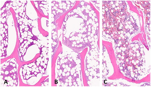
Figure 8. Transverse CT and MRI images of the L1 vertebra with anatomic right on the left side of the images. (A) CT image with dMWA applicator in the left aspect of the vertebral body and fiberoptic sensor in the right. (B) T1-weighted MRI image of L1 showing focal, semi-circular region of hyperintensity axial to the tract previously occupied by the dMWA applicator. (C) T2-weighted MRI image with fat saturation. Note the faint hyperintense rim along the peripheral aspect of the hyperintense region seen in the T1-weighted image (outlined by white arrows).
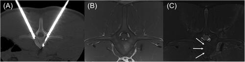
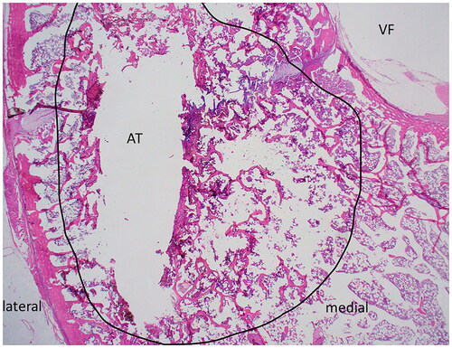
Figure 9. H&E stained slide of transverse section of ablated vertebrae L5. Medial and lateral margins are shown for orientation; the vertebral foramen (VF) and ablated tract (AT) are indicated. The region of thermal damage is circled in black.
Data availability statement
The data that support the findings of this study are available from the corresponding author upon reasonable request.