
Figures & data
Figure 1. Image profile of a 24-year-old patient with UAVF. (A) Ultrasound images of the patient with uterine arteriovenous fistula. (B) Grayscale ultrasound showed thickening of the myometrium, and no echo in local pipe-like/honeycomb shape. Color Doppler showed multicolored blood flow signals in the anechoic region. (C) The pre-HIFU MR image showed a lesion located at the bottom of the uterus. (D) The post-HIFU MR image showed no perfusion in the lesion located at the anterior wall of the uterus. (E) Ultrasound image of uterus 1 month after HIFU showed that the lesion reduced. (F) Ultrasound image of the uterus 12 month after HIFU showed good endometrial continuity and good triple line endometrial pattern.
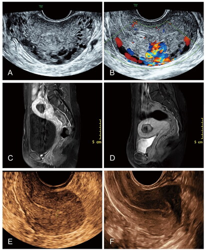
Figure 2. Image profile of a 33-year-old patient with UAVF. (A) Color Doppler ultrasonography of a longitudinal section of the patient’s uterus before HIFU treatment. (B) Color Doppler showed multicolored blood flow signals in the anechoic region. (C) The pre-HIFU MR image showed a lesion located at left anterior wall of the uterus. (D) The post-HIFU MR image showed no perfusion in the lesion located at the anterior wall of the uterus. (E) Ultrasound examination performed one month later showed no blood flow, with the lesion measuring 19 × 17 × 13 mm and a reduction rate of 57%. (F) Ultrasound image of the uterus 12 month after HIFU showed a relatively normal uterine cavity.
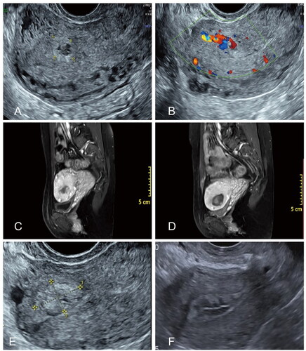
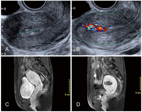
Figure 3. Image profile of a 32-year-old patient with UAVF. (A and B) Color Doppler ultrasonography of the patient’s uterus before HIFU treatment. (C) The pre-HIFU MR image showed a lesion located at the anterior wall of the uterus. (D) The post-HIFU MR showed non-perfusion in the lesion immediately after HIFU treatment.
Data availability statement
The data presented in this study are available on request from the corresponding authors.