
Figures & data
Figure 1. Endoscopic images of patients with VaIN I before and after treatment are shown in . (a) Represents the appearance of the lesion site before treatment when 3% glacial acetic acid was applied. (b) Shows the lesion site before treatment when Lugol’s iodine solution was applied for recurrent staining. (c) Displays the lesion site after 6 months reexamination following treatment when 3% glacial acetic acid was applied. Finally, (d) demonstrates the lesion site after half a year’s reexamination following treatment when Lugol’s iodine solution was applied. The blue arrow indicates the location of the lesion in the images.
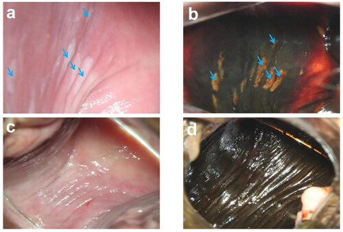
Figure 2. Endoscopic images of patients with vaginal stump VaIN I before and after treatment are presented in . (a) Shows the appearance of the lesion site before treatment when 3% glacial acetic acid was applied. (b) Displays the lesion site before treatment when Lugol’s iodine solution was applied for recurrent staining. After half a year’s reexamination following treatment, (c) exhibits the lesion site when 3% glacial acetic acid was applied. Finally, (d) demonstrates the lesion site after half a year’s reexamination following treatment when Lugol’s iodine solution was applied. The blue arrow indicates the location of the lesion in the images.
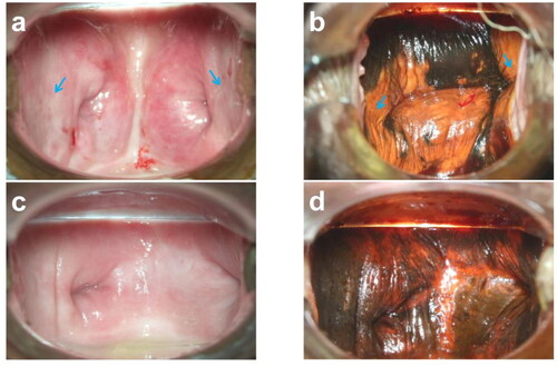
Table 1. Case data characteristics of VaIN patients.
Table 2. Effect of HIFU on VaIN.
Data availability statement
All data are made available by the authors on request without undue reservation. Data openly available in a public repository. The data that support the findings of this study are openly available in https://doi.org/10.6084/m9.figshare.24899691.v1