
Figures & data
Figure 1. Experimental design five treatment groups were used to investigate the effects of histotripsy treatment through gas blockage. From left: Control treatments without blockage, bowel positioned 0 cm above the agar phantom at both normal and double amplitude, and bowel positioned 1 and 2 cm above the phantom. The therapeutic beam path is denoted by the dotted lines. A 2.5 cm diameter spherical treatment (red circle) was prescribed in each phantom. Following histotripsy treatment, the bowel was inspected for gross and microscopic signs of damage.
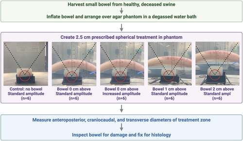
Figure 2. Experimental setup. A) the treatment head (pink arrow) of a clinical-grade histotripsy system was positioned in a degassed water bath above an agar phantom (white arrows) with gas-filled small bowel (yellow arrows) partially blocking the therapeutic beam path. B) Bowel was positioned 0 cm above the phantom (white arrow), shown in the sagittal plane. C) An image from the co-axially mounted diagnostic ultrasound transducer shows the bowel (yellow arrow) positioned above the phantom (white arrow) in the axial plane, which has been obscured by the intraluminal gas.
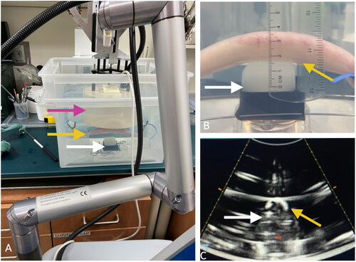
Figure 3. Treated Phantoms. Phantoms (4.6 cm × 4.6 cm × 4.6 cm) were sliced twice following treatment to facilitate measurements in both the axial and sagittal planes. A) The AP and TRANS diameters were visible following a slice in the axial plane, illustrated by the drawing on the right. B) The AP and CC diameters in the same phantom were visible in the sagittal plane.
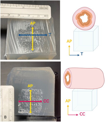
Figure 4. Computer simulation a ray-tracing approach was used to estimate the amount of the therapeutic transducer face (blue) blocked by gas-filled bowel (green) that extends in the CC direction. Yellow rays represent unobstructed therapeutic ultrasound that reaches the target while red rays represent blocked ultrasound intersecting with gas-filled bowel.
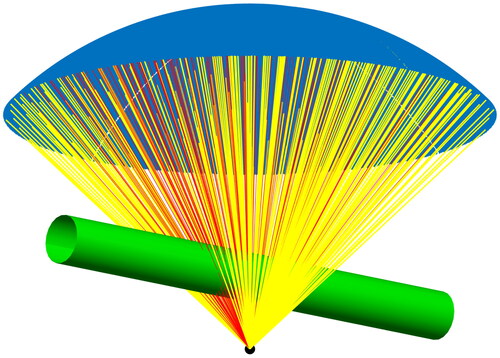
Figure 5. Gross and microscopic pathology. A) bivalved small bowel sectioned longitudinally. There were no signs of visible damage to the small bowel mucosa following treatment. B) Treated tissue taken from the center point of the gross sample shows intact mucosa, lamina propria, submucosa, and muscularis propria (20× magnification, H&E). C) untreated control tissue harvested from bowel outside of the ultrasound beam demonstrates no signs of damage (20× magnification, H&E).
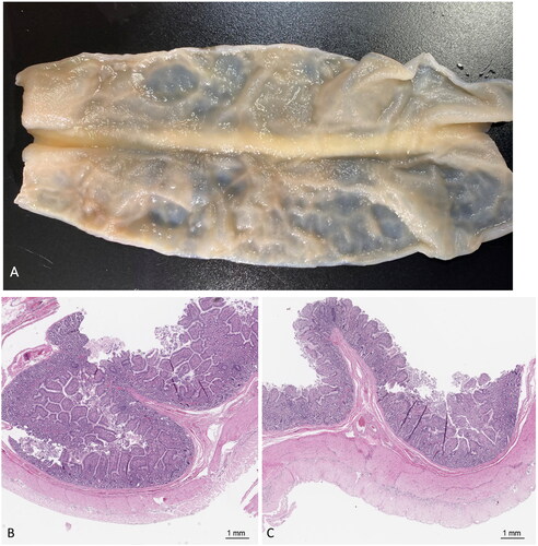
Table 1. Summary of histotripsy treatment volume.
Table 2. Summary of treatment amplitudes.