
Figures & data
Figure 1. Valve designs over the ages.
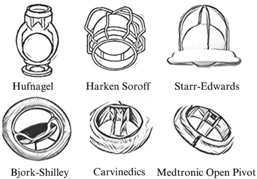
Figure 2. Carbomedics valve. Valve ring and leaf made of Si-doped pyrolytic carbon [Citation4].
![Figure 2. Carbomedics valve. Valve ring and leaf made of Si-doped pyrolytic carbon [Citation4].](/cms/asset/3d37554b-a5e8-4673-9266-fa889e0585a2/ysue_a_2238971_f0002_oc.jpg)
Figure 3. Platelet adhesion on the different phases after 120 minutes under static conditions. Image reproduced without modification from [Citation9].
![Figure 3. Platelet adhesion on the different phases after 120 minutes under static conditions. Image reproduced without modification from [Citation9].](/cms/asset/f82755e5-7ab3-4adc-9b6c-e51a3dd752f1/ysue_a_2238971_f0003_ob.jpg)
Figure 4. Thrombin generation of fresh human platelets after a 1-hour exposure. Image reproduced without modification from [Citation12].
![Figure 4. Thrombin generation of fresh human platelets after a 1-hour exposure. Image reproduced without modification from [Citation12].](/cms/asset/dcc33f5f-b875-4e42-978d-8ccf1d93d6f1/ysue_a_2238971_f0004_oc.jpg)
Figure 5. Blood droplet falling/bouncing on a pyrolytic carbon (PyC) leaflet (top) and a PyC leaflet with a hierarchical coating (bottom) from a height of 10 mm. Image reproduced without modification from [Citation16].
![Figure 5. Blood droplet falling/bouncing on a pyrolytic carbon (PyC) leaflet (top) and a PyC leaflet with a hierarchical coating (bottom) from a height of 10 mm. Image reproduced without modification from [Citation16].](/cms/asset/50628085-e816-43d3-a695-9081a4b9c196/ysue_a_2238971_f0005_ob.jpg)
Figure 6. (a) SEM image of the micro-structured cobblestone surface structure of rabbit valve and (b) magnified image of a single cobblestone. Image reproduced without modification from [Citation22] (c) Detailed heart valve structure. Image reproduced without modification from [Citation17].
![Figure 6. (a) SEM image of the micro-structured cobblestone surface structure of rabbit valve and (b) magnified image of a single cobblestone. Image reproduced without modification from [Citation22] (c) Detailed heart valve structure. Image reproduced without modification from [Citation17].](/cms/asset/fd5b303e-5e55-4a0a-9936-10572681d50f/ysue_a_2238971_f0006_oc.jpg)
Figure 7. The four surface textures formed on occluders subjected to ultra-short pulse laser and their water contact angles. Image adapted from [Citation23].
![Figure 7. The four surface textures formed on occluders subjected to ultra-short pulse laser and their water contact angles. Image adapted from [Citation23].](/cms/asset/6ae345bf-b383-40a0-a2a9-7f3e363e1d40/ysue_a_2238971_f0007_ob.jpg)