5,763
Views
111
CrossRef citations to date
0
Altmetric
Original Articles

Can tDCS enhance treatment of aphasia after stroke?

&
Pages 1169-1191 | Published online: 03 Nov 2011

Figures & data

TABLE 1 tDCS and language performance in post-stroke aphasia

Figure 1. Summary schematic representation of potential tDCS effects in language performance in relation to normal re-learning after single and repeated interventions protocols (related to ). Solid green line represents normal re-learning curve. Solid vertical red line indicates the potential percentage change in performance that may occur after a single intervention of tDCS. Dashed red line indicates potential performance change over 5 days from repeated tDCS delivered concurrently with treatment. Normal and tDCS-enhanced learning share an equivalent learning profile, however resulting gains post-tDCS may be modified by approximately 25%, with the effects of tDCS persisting for up to 3 weeks. To view this figure in colour, please see the online issue of the Journal.
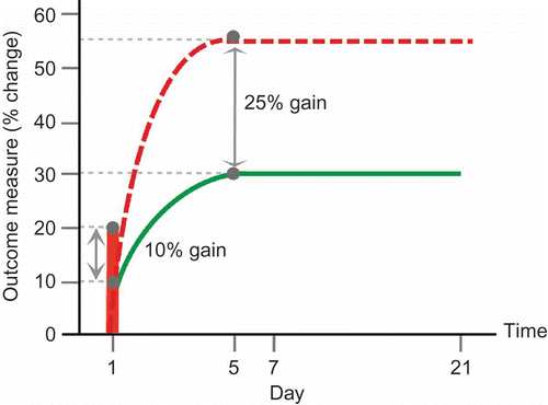
Figure 1. Summary schematic representation of potential tDCS effects in language performance in relation to normal re-learning after single and repeated interventions protocols (related to Table 1). Solid green line represents normal re-learning curve. Solid vertical red line indicates the potential percentage change in performance that may occur after a single intervention of tDCS. Dashed red line indicates potential performance change over 5 days from repeated tDCS delivered concurrently with treatment. Normal and tDCS-enhanced learning share an equivalent learning profile, however resulting gains post-tDCS may be modified by approximately 25%, with the effects of tDCS persisting for up to 3 weeks. To view this figure in colour, please see the online issue of the Journal.

Figure 2. Illustration of potential target sites of stimulation to facilitate naming performance in relation to structural brain damage and the normal naming network in three different patients. The top panel illustrates the extensive bilateral fronto-temporal naming network found in an fMRI study of healthy older particpants (cf. Holland et al., Citation2011). Activation is overlaid onto a rendered cortical surface from SPM8. In the lower panels of the figure we overlay the normal pattern of brain activation onto three chronic aphasic stroke patients' individual structural MRI scans. The aim here is to illustrate from left to right for each patient: (1) their structural brain damage within the left hemisphere, (2) the normal naming network overlaid onto their individual brain scan to show the relationship between the lesion damage to the naming network and the target stimulation site (red cross), and (3) the stimulation sites feasible for each patient depending on the proposed target site. Panel A highlights, in two chronic aphasic stroke patients, that structurally intact regions of cortex within the lesioned left hemisphere may serve as potential candidate sites for anodal stimulation to facilitate treatment of anomia in (Ai) Broca's area and (Aii) Wernicke's area. Panel B highlights that when the lesion is has damaged relevant cortices in the left hemisphere perilesional stimulation may not be possible. Therefore, for the two patients illustrated, facilitation of the contralesional hemisphere may be the optimal approach to aid recovery: (Bi) right homologue to Broca's area and (Bii) Wernicke's area. Although not illustrated here, in cases where patients have suffered a large MCA infarct affecting the whole of the left hemisphere and resulted in extremely limited perilesional tissue, stimulation of the contralesional hemisphere would be the sole option. Furthermore, studies of aphasia recovery have successfully used anodal stimulation of the left perilesional hemisphere to elicit positive behavioural outcomes, however the effects of anodal or cathodal stimulation of the right contralesional hemisphere are, as yet unknown, and will be dependent on the theoretical hypotheses of the research. To view this figure in colour, please see the online issue of the Journal.
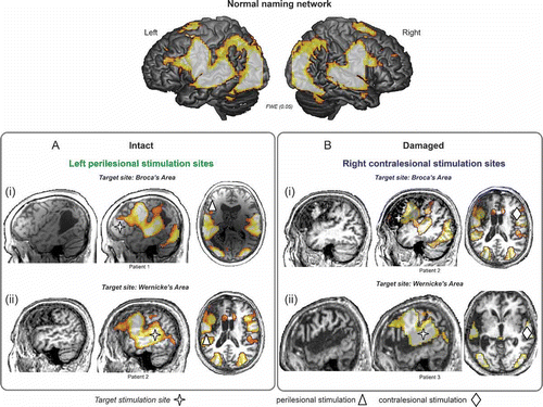
Figure 2. Illustration of potential target sites of stimulation to facilitate naming performance in relation to structural brain damage and the normal naming network in three different patients. The top panel illustrates the extensive bilateral fronto-temporal naming network found in an fMRI study of healthy older particpants (cf. Holland et al., Citation2011). Activation is overlaid onto a rendered cortical surface from SPM8. In the lower panels of the figure we overlay the normal pattern of brain activation onto three chronic aphasic stroke patients' individual structural MRI scans. The aim here is to illustrate from left to right for each patient: (1) their structural brain damage within the left hemisphere, (2) the normal naming network overlaid onto their individual brain scan to show the relationship between the lesion damage to the naming network and the target stimulation site (red cross), and (3) the stimulation sites feasible for each patient depending on the proposed target site. Panel A highlights, in two chronic aphasic stroke patients, that structurally intact regions of cortex within the lesioned left hemisphere may serve as potential candidate sites for anodal stimulation to facilitate treatment of anomia in (Ai) Broca's area and (Aii) Wernicke's area. Panel B highlights that when the lesion is has damaged relevant cortices in the left hemisphere perilesional stimulation may not be possible. Therefore, for the two patients illustrated, facilitation of the contralesional hemisphere may be the optimal approach to aid recovery: (Bi) right homologue to Broca's area and (Bii) Wernicke's area. Although not illustrated here, in cases where patients have suffered a large MCA infarct affecting the whole of the left hemisphere and resulted in extremely limited perilesional tissue, stimulation of the contralesional hemisphere would be the sole option. Furthermore, studies of aphasia recovery have successfully used anodal stimulation of the left perilesional hemisphere to elicit positive behavioural outcomes, however the effects of anodal or cathodal stimulation of the right contralesional hemisphere are, as yet unknown, and will be dependent on the theoretical hypotheses of the research. To view this figure in colour, please see the online issue of the Journal.