1,822
Views
21
CrossRef citations to date
0
Altmetric
Review

Dopaminergic therapy in aphasia

&

Figures & data

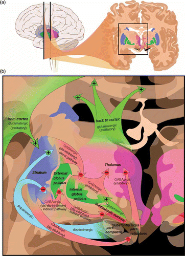
Figure 1. (a) The two main dopaminergic nuclei are close to each other in the midbrain with the substantia nigra projecting to the striatum and the ventral tegmental area projecting to both the nucleus accumbens (part of the ventral striatum) and the frontal cortex. (b) The nigrostriatal pathway affects cortical function indirectly through a series of cortico-basal ganglia circuits. The substantia nigra (pars compacta) projects to the striatum (caudate and putamen in blue). The main output of the striatum is to the globus pallidus (green) and thence to the thalamus (pink) and cortex (green arrows). The cortex feeds back to the striatum (green arrows) to close the loop on the cortico-basal ganglia circuits. There are also dopaminergic receptors on many cortical neurons (not shown). http://en.wikipedia.org/wiki/File:Basal_ganglia_circuits.svg. Mikael Häggström, based on images by Andrew Gillies.

Figure 1. (a) The two main dopaminergic nuclei are close to each other in the midbrain with the substantia nigra projecting to the striatum and the ventral tegmental area projecting to both the nucleus accumbens (part of the ventral striatum) and the frontal cortex. (b) The nigrostriatal pathway affects cortical function indirectly through a series of cortico-basal ganglia circuits. The substantia nigra (pars compacta) projects to the striatum (caudate and putamen in blue). The main output of the striatum is to the globus pallidus (green) and thence to the thalamus (pink) and cortex (green arrows). The cortex feeds back to the striatum (green arrows) to close the loop on the cortico-basal ganglia circuits. There are also dopaminergic receptors on many cortical neurons (not shown). http://en.wikipedia.org/wiki/File:Basal_ganglia_circuits.svg. Mikael Häggström, based on images by Andrew Gillies.

TABLE 1 Summary of the 15 studies covered by this review