Figures & data
Figure 1. Computed tomography of the patient’s brain.
Left thalamic hemorrhage with ventricle rupture as seen immediately after the hemorrhage (day 1), and the same area at year 2 post onset. The lesion mainly involved middle-to-posterior parts of the thalamus, including the pulvinar, lateral posterior, centromedian, and reticular nuclei.
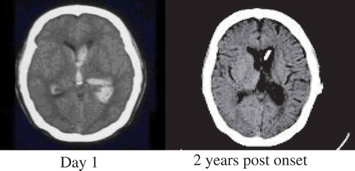
Table 1. Results of neuropsychological assessments.
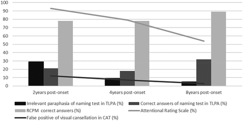
Figure 2. Changes in linguistic and neuropsychological function over the course of the study. The decline in the patient’s irrelevant paraphasias correlated with improved attentional function but not with intelligence or word-retrieval ability. CAT = Clinical Attention Assessment Test; RCPM = Raven’s Colored Progressive Matrices; TLPA = Test of Lexical Processing in Aphasia.