
Figures & data
Figure 1. Pre- and postoperative post-contrast T1-weighted MRI scans in the axial and sagittal plane in patients A1, B2, C3, D4 and E5. Pre = preoperative. Post = postoperative
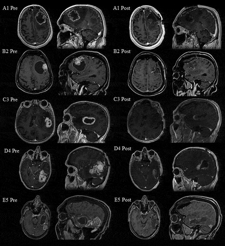
Table 1. Demographic and clinical characteristics.
Table 2. Neurolinguistic assessment
Table 3. Pre- and postoperative neurolinguistic test-protocol (raw score/z-score).
Supplemental material