
Figures & data
Figure 1. MRI’s made during follow-up. I: First MRI (2014) revealing a suprasellar hyperdense and an infrasellar hypodense lesion and a nasal tumour. II: At follow up (2015) the difference in density is less clear. III Directly preoperative MRI (2016) showing an isodense lesion.
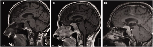
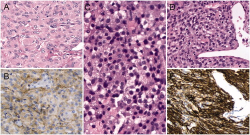
Figure 2. Histological examination reveals a meningioma (A) with positive staining for Epithelial Membrane Antigen (B), a pituitary adenoma (C), and a glomangiopericytoma (D) with positive staining for SMA (E).
Supplemental material