Figures & data
Table 1. Diagnosis of schwannomatosis based on molecular and/or clinical diagnostic criteria according to Plotkin et al.Citation6 and Pstrow et al.Citation11
Figure 1. (A) Low power view of a schwannoma, showing an encapsulated spindle cell lesion and vessels with hyaline walls. (B) Presence of diffuse S-100 staining.
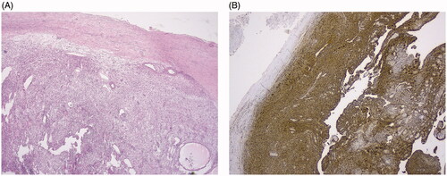
Figure 2. (A) T1-weighted MRI scan with contrast demonstrating the contrast enhanced schwannomas. (B) T1-weighted MRI scan without contrast.
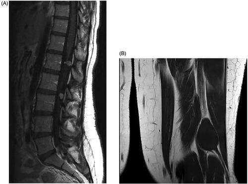
Figure 3. Hyperintense lesions on T2-weighted MRI scan without contrast.
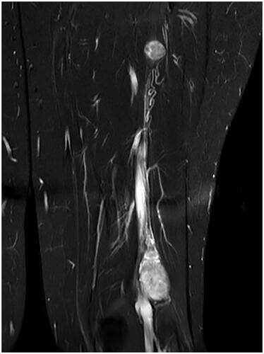
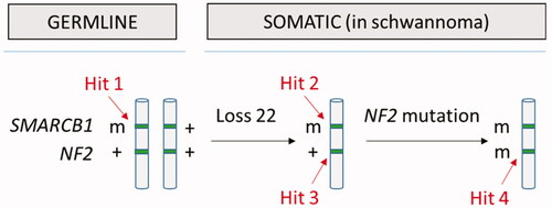
Figure 4. The four-hit, three-step model for tumorigenesis in schwannomatosis (here shown for SMARCB1): germline mutation in SMARCB1 (hit 1), loss of a region of chromosome 22 (hit 2 and 3) and pathogenic mutation in remaining WT NF2 allele (m = pathogenic mutation; + = WT). The same mechanism has also been demonstrated for germline LZTR1 mutations.