
Figures & data
Figure 1. Preoperative sagittal (A), axial (B), and coronal (C) T1-weighted MRI with gadolinium depicting a large OGM with mass effect on the surrounding frontal lobes. The patient underwent a right LSO approach with Simpson grade II resection of the tumor as demonstrated on the postoperative sagittal (D), axial (E), and coronal (F) T1-weighted postcontrast MRI.
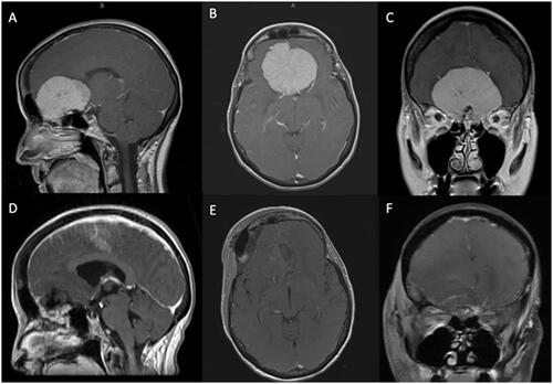
Table 1. Patients and tumor characteristics.
Table 2. Patients’ outcomes following LSO approach.
Table 3. Follow-up and adjuvant therapy.