Figures & data
Table 1. Comparison of morphological features of Entomoneis annagodhei to similar species.
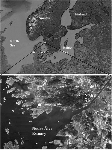
Fig. 1. Map indicating sampling site, Nodre Älv Estuary, located at the west coast of Sweden.
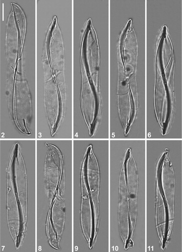
Figs 2–11. LM micrographs of holotype specimen of Entomoneis annagodhei sp. nov. at different foci showing general size diminution of valves. Note oblique transapical fascia (Figs 2–6; 8 and 11) and central area of the raphe canal with lanceolate slit-like opening (Figs 7, 9 and 10). Scale bar = 10 µm.
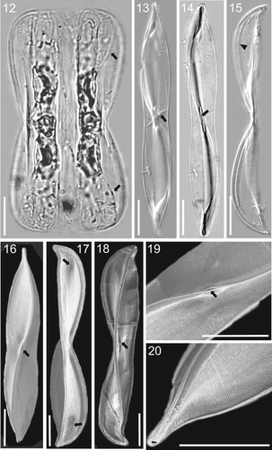
Figs 12–20. LM (Figs 12–15) and SEM (Figs 16–20) micrographs of holotype specimen of Entomoneis annagodhei sp. nov. Fig. 12. Panduriform cell containing two plate plastids and numerous intercalary bands and straight to slightly arcuate transition between valve body and keel (arrows). Figs 13–14. Linear to lanceolate valves of same specimen observed with DIC (Fig. 13) and without (Fig. 14) showing slightly concave valve margins at the centre of valve length and slightly protracted apices. Oblique transapical fascia (Fig. 13, arrow). Central area of raphe canal showing lanceolate slit-like opening (Fig. 14, arrow). Fig. 15. Girdle view of the valve with acuminate apices and pronounced narrow raphe canal separated with raphe fibulae in form of a line (arrowhead). Fig. 16. Valve view showing sigmoid raphe canal, dense transapical striae and lanceolate slit-like opening at the central raphe canal area (arrow). Fig. 17. Girdle view of the valve showing depressions at the transition between valve body and keel (arrows). Fig. 18. Girdle view of the valve showing inner structure and oblique transapical fascia (arrow). Fig. 19. Details of Fig. 16, showing fine valve striation and lanceolate slit-like opening (arrow). Fig. 20. Acuminate valve apex showing hooked terminal raphe fissure (arrow). Scale bar = 10 µm.
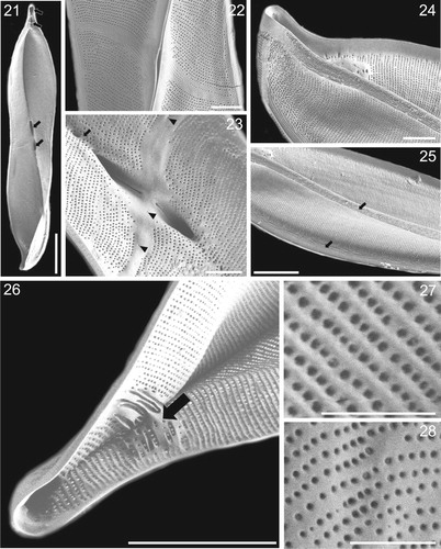
Figs 21–28. SEM microghraphs of Entomoneis annagodhei sp. nov. Fig. 21. Internal valve view showing apical and central sub-compartments (arrows). Fig. 22. External valve view showing oblique transapical fascia and lanceolate slit-like opening of deep raphe canal. Fig. 23. Internal view of central valve area showing oblique transapical fascia with few isolated areolae (arrowheads), central raphe fissures opening into fascia and dense raphe fibulae forming a line (arrow). Fig. 24. External valve view showing acuminate apex and dense smooth and parallel transapical striae and virgae. Fig. 25. External valve view showing linear to undulate external ridges running more or less parallel, and adjacent to the raphe (arrows). Fig. 26. Internal valve view of the apical sub-compartments and irregularly shaped and thickened basal fibulae (arrow). Figs 27–28. External and internal valve view with areola details. Scale bar = 10 µm (Fig. 21), 2 µm (Figs. 22–24), 5 µm (Figs. 25–26), 1 µm (Figs. 27–28).