
Figures & data
Table 1. Sources of samples from Yap (Y) and Palau (PW) used in this study.
Figs 1–8. Gomphotheca marciae n. sp., whole valves and external aspect (LM and SEM). Fig. 1. Preserved cell showing plastids. Fig. 2. Cleaned valve in LM (isotype). Figs 3, 4. Whole valves in SEM, external and internal aspects. Fig. 5. Detail of plastids. Figs 6, 7. Apical and basal poles, external aspect. Fig. 8. External detail showing raphe slit with lateral depressions along each side. Scale bars: Figs 1–4 = 50 µm, Fig. 5 = 10 µm, Figs 6–8 = 2 µm.
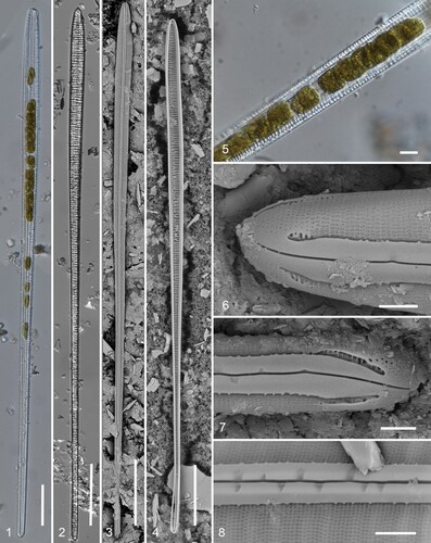
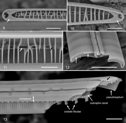
Figs 9–13. Gomphotheca marciae n. sp., internal aspect and valve structure (SEM). Figs 9, 10. Internal details of apical and basal poles showing pseudosepta (arrows). Fig. 11. Internal detail showing costate fibulae; white arrow points to row of pores in subraphe canal, black arrow to covering of canal between some fibulae. Interior of valvocopula seen across bottom of image. Fig. 12. Broken valve showing cross-sectional shape. Notice the pores passing through the attachment of the costate fibulae (long arrow) vs. basal silica layer (short arrow). Fig. 13. Valve broken near basal pole in oblique view, showing conopeum with pores beneath it, raphe slit with lateral depressions, subraphe canal, portion of pseudoseptum, and complexity of valve structure at the pole (black arrow). Scale bars: Figs 9–12 = 5 µm, Fig. 13 = 2 µm.
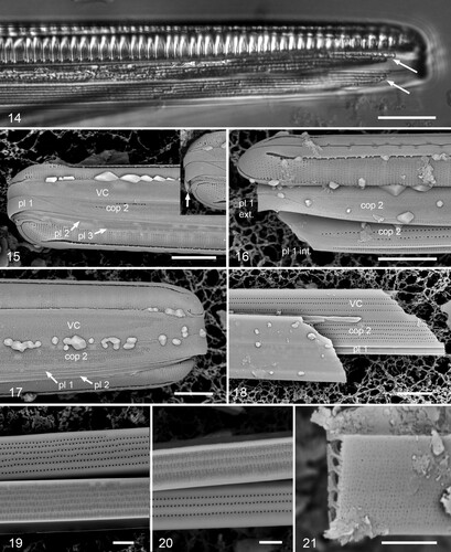
Figs 14–21. Gomphotheca marciae n. sp., girdle bands (LM and SEM). Fig. 14. Girdle view in LM showing valve with two attached copulae, arrows indicating ends of the pores/longitudinal chambers. Figs 15, 16. Whole mounts in SEM showing basal poles, with and without the hypotheca; copulae and pleurae labeled (VC = valvocopula, cop 2 = second copula, pl 1 = first pleura, etc.). In Fig. 15, pl 1 is intact and shows the wide apical cap that fits against cop 2, and a third pleura can be seen; in Fig. 16, the apex of the cingulum has broken off, revealing the internal relationship between cop 2 and pl 1. Inset in Fig. 15 (same scale) shows pole with cracked pl 1, showing thickness at apex (arrow). Fig. 17. Whole mount showing apical pole; in this image the external poration of the girdle bands is very clear. Fig. 18. Broken cingulum showing especially in internal aspect of valvocopula (VC), second copula (cop 2) and first pleura (pl 1). Figs 19, 20. Portions of second copula at two distances from apex showing inner and outer poration and reduction in rows from six to three toward base. Fig. 21. Broken second copula showing longitudinal canals. Scale bars: Fig. 14 = 10 µm, Figs 15–18 = 5 µm, Figs 19–21 = 2 µm.
Figs 22–26. Gomphotheca marciae n. sp., copulae (SEM). Figs 22, 23. Basal and apical details of valvocopula in situ adjacent to the valve, showing that this band is closed and showing overlap of silica flaps on valvocopula with pseudoseptum on valve. Fig. 24. Oblique view of valvocopula showing silica flaps and ruffled groove of pars interior. Figs 25, 26. Open (basal) ends of a second copula showing external and internal poration. Scale bars 5 µm.
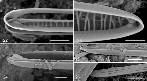
Table 2. Comparison of the morphology of Gomphotheca spp.