
Figures & data
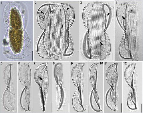
Figures 1–12. LM micrographs of Entomoneis grisslehamnensis sp. nov. at different focal planes. Fig. 1. Live cell in girdle view showing panduriform frustule and plate-like plastid. Figs 2–4. Panduriform frustule with elevated virgae on valve body (arrows), basal fibulae forming arcuate transition between bulged valve body and flattened wing (arrowheads) and numerous crossed girdle bands. Figs 5–6. Valves in girdle view showing scalpelliform apices. Figs 7–8. Apically torsioned valves. Figs 9–12. Valves in girdle view with pronounces raphe fibulae and elevated virgae on valve body. Scale bar = 10 µm.
Figures 13–20. SEM micrographs of Entomoneis grisslehamnensis sp. nov. Fig. 13. Frustule in girdle view. Figs 14–15. Valves in girdle view with girdle band details showing bulged valve body and flattened wings with pronounced elevated virgae and silicified rims on wings. Fig. 16. Valve in girdle view with arcuate transition between bulged valve body and flattened wing. Figs 17–18. Valves in valve view with sigmoid raphe canal on elevated keel. Fig. 19. Details of proximal raphe endings in central node. Fig. 20. Scalpelliform valve apex.
Figures 21–27. SEM and TEM micrographs of Entomoneis grisslehamnensis sp. nov. Figs 21, 22, 24 and 26. SEM; Figs 23, 25, 27. TEM. Fig. 21. Distal raphe ending (arrow) viewed from inner valve. Fig. 22. Tilted valve apex showing elevated virgae, thicker at the valve margin. Fig. 23. Elevated wing with radial striae appearing decussate. Fig. 24. Bulged valve body and flattened wing with silicified rims around areolae; specific row of areolae on raphe-canal. Figs 25–27. Details of quadrangular areolae perforated with finely perforated hymen.
Figures 28–33. LM micrographs of Entomoneis paludosa at different focal planes. Fig. 28. Panduriform frustule in girdle view with sinusoid transition between valve body and keel, pronounced raphe canal and fibulae and numerous crossed girdle bands. Figs 29–33. Different valve sizes viewed in girdle view showing differences in sinusoid curvature of transition between valve body and keel. Scale bar = 10 µm.
Figures 34–40. SEM and TEM micrographs of Entomoneis paludosa. Figs 34, 35, 36, 38 and 40. SEM; Figs 37 and 39. TEM. Fig. 34. Frustule in girdle view showing sinusoid transition between valve body and keel, pronounced raphe canal and numerous intercalary bands. Fig. 35. Linear to lanceolate valve with three distinctive bulges (arrows). Figs 36 and 37. Valves in girdle view with distinctive sinusoid transition from valve body to keel viewed in SEM (Fig. 36) and TEM (Fig. 37). Figs 38 and 40. Close-up of wing showing regular areolar striae and comb-like structure made from hymenated striae. Fig. 39. Protracted valve apex with areolae details.
Figures 41–50. SEM and TEM micrographs of Entomoneis paludosa. Figs 41 and 42. SEM; Figs 43–50. TEM. Fig. 41. Tilted central valve showing flattened wing at the central node ang bulged valve body. Fig. 42. Proximal raphe endings and raphe canal areolae. Fig. 43. Details of striation in the wing and valve body. Figs 44 and 45. Close-up of raphe canal striae, raphe fibulae (RF) and keel fibulae (KF) with comb-like striae continuing towards transition between keel and valve body (Fig. 44.). Figs 46–49. Details of perforations in elliptical areolae and comb-like hymenated striae. Fig. 50. Valve margin with valvocopulae ultrastructure showing smaller advalvar and larger abvalvar elongated areolae.
Table 1. Morphological features of Entomoneis grisslehamnensis sp. nov. compared to similar species: E. decussata, E. aequabilis, E. paludosa, E. tenera and E. punctulata.