Figures & data
Figure 1. Collecting site locations within the GLSM's watershed.
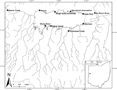
Table 1. Physiochemical parameters and habitat characterization for sites within the GLSM watershed. All sites were sampled between 1 June 2015 and 1 July 2015.
Figure 2. Location of 18 manually set landmarks used for morphological analysis of Lepomis macrochirus: 1 tip of snout; 2 nuchal hump (follows angle of operculum to body); 3 anterior insertion of dorsal fin; 4 posterior insertion of dorsal fin; 5 insertion of last dorsal ray on caudal fin; 6 insertion of last ventral ray of caudal fin; 7 posterior insertion of anal fin; 8 anterior insertion of anal fin; 9 insertion of pelvic fin; 10 gill juncture; 11 midpoint of eye; 12 insertion of pectoral fin; 13 ventral insertion of opercular flap; 14 dorsal insertion of opercular flap; and 15 posterior edge of opercular flap. Landmarks numbered 1, 16, 17, and 18 were used to digitally unbend each specimen and thereby not included in the analyses.
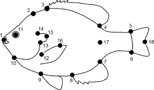
Table 2. MANOVA results of relative warp analyses of all individuals collected from the GLSM watershed. Length is the primary predictor of morphology in Bluegill.
Figure 3. Overall visualizations for morphological variation in the GLSM watershed area.
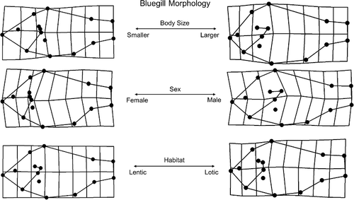
Table 3. GLM results of relative warp analyses of all individuals collected from the GLSM watershed.
Figure 4. RWA for three primary axes which explained 65 percent of morphological variation in the GLSM watershed area, including significant relationships with parent terms as shown in general linear models.
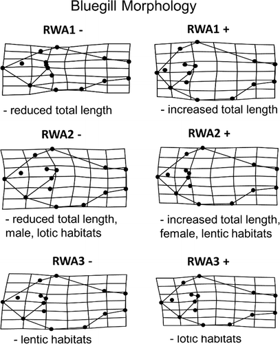
Figure 5. Scatterplot of RWA1 and RWA2 for both male and female individuals in the GLSM watershed. Closed circles indicate females and open circles indicate males.
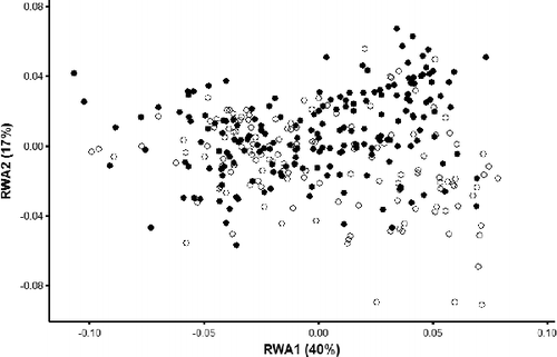
Figure 6. Scatterplot of RWA 1 and total length with regressions for Bluegill. Closed circles and solid regression line indicate females and open circles and dashed regression line indicate males.
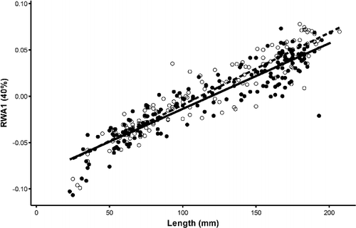
Figure 7. Scatterplot of RWA2 and total length (mm) with regressions for Bluegill individuals. Closed circles and solid regression line indicate females and open circles and dashed regression line indicate males.
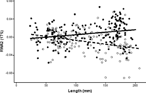
Figure 8. Scatterplot of RWA2 and total length (mm) with regressions for Bluegill individuals. Closed circles and solid regression line indicate individuals from lentic habitats and open circles and dashed regression line indicates individuals from lotic habitats.
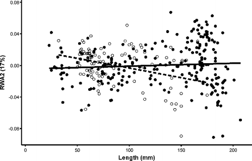
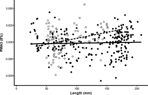
Figure 9. Scatterplot of RWA3 and total length (mm) with regressions for Bluegill individuals. Closed circles and solid regression line indicate individuals from lentic habitats and open circles and dashed regression line indicates individuals from lotic habitats.