
Figures & data
Table 1a. Number and characteristics of the subjects.
Table 1b. Check list of objective finding scores for superior limbic keratoconjunctivitis.
Table 2. The clinical scores and SHI of patients in the SLK and SS groups categorized according to age, sex, and diagnosis.
Figure 1. Anterior photographs of a representative patient and the relationship between the clinical scores and superior hyperemia index (SHI).
a, b, and c: Slit-lamp photographs of the patient with superior limbic keratoconjunctivitis show that the patient’s eye scores one point each for papillary formation of the superior palpebral conjunctiva, superior limbal swelling and hyperemia, and superior corneal epitheliopathy.
d, e, and f: Relationship between the SHI and papillary formation of the superior palpebral conjunctiva (d), superior limbal hyperemia (e) and swelling, and superior corneal epitheliopathy (f). The SHI shows a tendency to indicate the severity of the superior limbal hyperemia and swelling score.
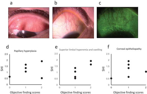
Table 3. Diagnostic usefulness of the superior hyperemia index (SHI).