Figures & data
Figure 1. Effects of 17β-estradiol (E2) on gene expression levels of superoxide dismutases, SOD1, SOD2, and SOD3, in human lens epithelial cells (HLECs).
No significant changes were seen in the gene expression levels of SOD1, SOD2, or SOD3 after exposure to E2 for 1.5 h or 24 h. Relative gene expression data normalized to reference genes, RPLP0 and PPIA, and shown as fold change compared to control cells (0 µM E2). Each dot in the same color represents samples from the same cell culture, and the three different colors represent the three different cell cultures (n = 3).
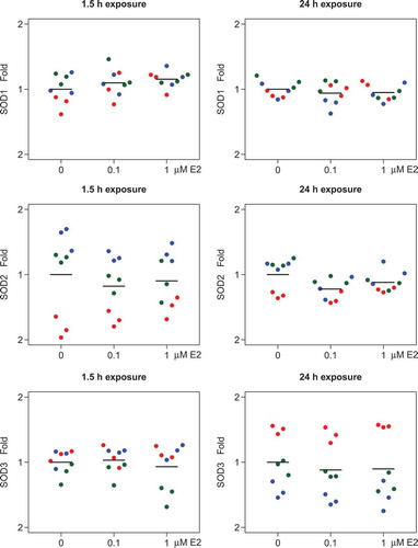
Figure 2. Effects of 17β-estradiol (E2) on superoxide dismutase protein expression levels in human lens epithelial cells (HLECs).
No significant changes were seen in SOD-1 (A, B) or SOD-2 (C, D) expression levels after exposure to E2 for 1.5 h or 24 h. Data presented from densitometric analyses of Western blot bands normalized to β-actin (n = 3) and shown as mean ± SD.
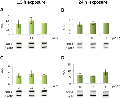
Figure 3. Immunolocalization of superoxide dismutases, SOD-1 and SOD-2, in human lens epithelial cells (HLECs) after exposure to 17β-estradiol (E2) for 1.5 h.
Nuclear morphology is shown with Hoechst 33342 and MitoTracker Deep Red FM was used for mitochondrial localization. Cells were labeled with antibodies against SOD-1 and SOD-2 and visualized by Alexa Fluor 488. Immunolabeling in the cytosol and nucleus was seen with SOD-1 antibody (A, B, C) Mitochondrial localization was evident by colocalization of SOD-2 with MitoTracker (D, E, F). Original magnification: 1000×. Scale bar: 20 µm.
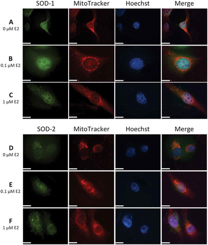
Figure 4. Increased superoxide dismutase (SOD) activity levels after exposure to 17β-estradiol (E2) in human lens epithelial cells (HLECs).
Significant increase in activity levels after exposure to 0.1 µM and 1 µM E2 for 1.5 h (A). No significant changes were seen in activity after 24 h exposure (B) Data presented as SOD units related to protein concentration (U/mg) shown as mean ± SD. Asterisks indicate statistical significance p ≤ 0.05 for comparison with control cells (0 µM E2).
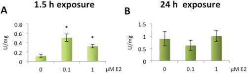
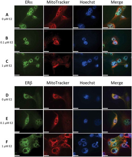
Figure 5. Altered protein expression levels of estrogen receptors (ERα and ERβ) after exposure to 17β-estradiol (E2) in human lens epithelial cells (HLECs)..
Decrease in ERα expression levels after exposure to 1 µM E2 for 1.5 h and 24 h (A, B). Elevated ERβ expression levels after exposure to 0.1 µM and 1 µM E2 for 1.5 h (C) and 1 µM E2 for 24 h (D). Data presented from densitometric analyses of Western blot bands normalized to β-actin (n = 3) shown as mean ± SD. Asterisks indicate statistical significance p ≤ 0.05 for comparison with control cells (0 µM E2).
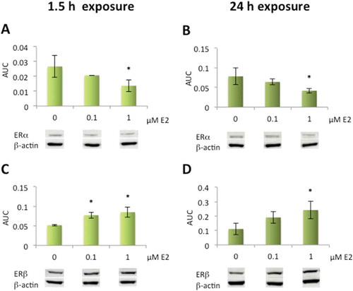
Figure 6. Immunolocalization of estrogen receptors, ERα and ERβ, in human lens epithelial cells (HLECs) after exposure to 17β-estradiol (E2) for 1.5 h.
Nuclear morphology is shown with Hoechst 33342, and MitoTracker Deep Red FM was used for mitochondrial localization. Cells were also labeled with antibodies against ERα and ERβ and visualized by Alexa Fluor 488. Images show ERα (A, B, C) and ERβ (D, E, F) localization in nucleus. Mitochondrial localization of ERβ is evident by the colocalization with MitoTracker, showing a more intense yellow color in merged images (D, E, F) compared to no colocaliztion with ERα (A,B,C). The immunolabeling of ERβ increased slightly at the highest E2 concentration, 1 µM (F). Original magnification: 1000×. Scale bar: 20 µm.