
Figures & data
Figure 1. The graphical presentation of the refractive and visual outcomes: (A) the cumulative logMAR visual acuity; (B) the postoperative UDVA versus the preoperative CDVA; (C) the postoperative SE refraction; (D) the postoperative refractive cylinder; (E) the postoperative refractive sphere; (F) the scatterplot of the attempted preoperative SE refraction versus the achieved postoperative SE refraction; (G) the stability of the postoperative SE refraction; and (H) the stability of the postoperative refractive cylinder.
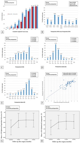
Table 1. CXL-Plus preoperative and postoperative data summary of the results during the follow-up period.
Table 2. Summary of the preoperative and postoperative data of a 14-year-old female patient.
Figure 2. Corneal topography of a 14-year-old female patient with KC: (A) preoperatively; (B) at the 6th follow-up month postoperatively with an improvement in the K readings; and at the 12th (C) and 18th (D) follow-up month postoperatively with stabile K readings.
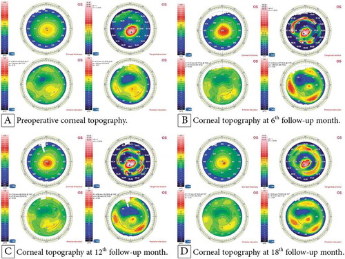
Figure 3. Corneal topography of a 9-year-old male patient with KC: (A) preoperatively; (B) at the 6th follow-up month postoperatively; (C) at the 12th follow-up month postoperatively; and (D) at the 18th follow-up month postoperatively.
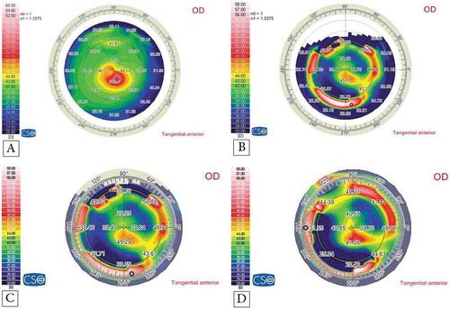
Figure 4. Corneal topography of a 17-year-old female patient with KC: (A) preoperatively; (B) at the 6th follow-up month postoperatively; (C) at the 12th follow-up month postoperatively; and (D) at the 18th follow-up month postoperatively.
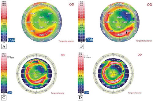
Table 3. Visual, topographic, and refractive pathways of the eight eyes with complications preoperatively and after 18 months.