2,408
Views
47
CrossRef citations to date
0
Altmetric
ARTICLES

A Late Cretaceous (Maastrichtian) snake assemblage from the Maevarano Formation, Mahajanga Basin, Madagascar

, , &
Pages 109-138 | Received 05 Jan 2009, Accepted 15 Apr 2009, Published online: 28 Jan 2010

Figures & data

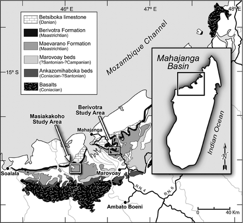
FIGURE 1 Late Cretaceous and Paleocene strata of the Mahajanga Basin, northwestern Madagascar. The Berivotra and Masiakakoho study areas are indicated by rectangular outlines.

FIGURE 1 Late Cretaceous and Paleocene strata of the Mahajanga Basin, northwestern Madagascar. The Berivotra and Masiakakoho study areas are indicated by rectangular outlines.

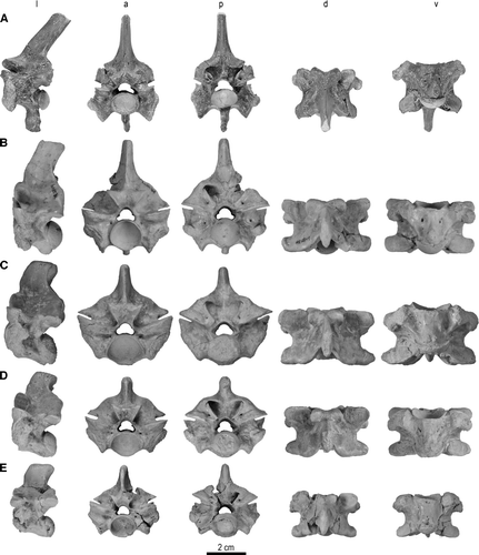
FIGURE 2 Trunk vertebrae of Madtsoia madagascariensis from the Late Cretaceous of Madagascar in l, lateral; a, anterior; p, posterior; d, dorsal; and v, ventral views. A, vertebra from anterior part of anterior trunk region with well-developed hypapophysis, FMNH PR 2546; B, vertebra from posterior part of anterior trunk region, FMNH PR 2549; C, mid-trunk vertebra, FMNH PR 2551; D, vertebra from anterior part of posterior trunk region, FMNH PR 2554; E, vertebra from middle part of posterior trunk region, FMNH PR 2555.

FIGURE 2 Trunk vertebrae of Madtsoia madagascariensis from the Late Cretaceous of Madagascar in l, lateral; a, anterior; p, posterior; d, dorsal; and v, ventral views. A, vertebra from anterior part of anterior trunk region with well-developed hypapophysis, FMNH PR 2546; B, vertebra from posterior part of anterior trunk region, FMNH PR 2549; C, mid-trunk vertebra, FMNH PR 2551; D, vertebra from anterior part of posterior trunk region, FMNH PR 2554; E, vertebra from middle part of posterior trunk region, FMNH PR 2555.

FIGURE 3 Cloacal and postcloacal vertebrae of Madtsoia madagascariensis from the Late Cretaceous of Madagascar in l, lateral; a, anterior; p, posterior; d, dorsal; and v, ventral views. A, cloacal vertebra, FMNH PR 2556; B, postcloacal vertebra, FMNH PR 2557. Articular facets on ventral aspect of postcloacal vertebra for chevron bone enlarged at bottom right.

FIGURE 3 Cloacal and postcloacal vertebrae of Madtsoia madagascariensis from the Late Cretaceous of Madagascar in l, lateral; a, anterior; p, posterior; d, dorsal; and v, ventral views. A, cloacal vertebra, FMNH PR 2556; B, postcloacal vertebra, FMNH PR 2557. Articular facets on ventral aspect of postcloacal vertebra for chevron bone enlarged at bottom right.

FIGURE 4 Ribs and rib fragments of Madtsoia madagascariensis from the Late Cretaceous of Madagascar. A, anterior; and B, posterior views of UA 9764, nearly complete left rib exhibiting a pathological lesion (indicated by arrows). C, anterior; D, posterior; and I, stereophotographic proximal views of UA 9746, proximal half of right rib (reversed to facilitate comparison). E, anterior; F, posterior; and J, stereophotographic proximal views of FMNH PR 2571, proximal fragment of left rib. G, anterior; and H, posterior views of UA 9763, nearly complete left rib.

FIGURE 4 Ribs and rib fragments of Madtsoia madagascariensis from the Late Cretaceous of Madagascar. A, anterior; and B, posterior views of UA 9764, nearly complete left rib exhibiting a pathological lesion (indicated by arrows). C, anterior; D, posterior; and I, stereophotographic proximal views of UA 9746, proximal half of right rib (reversed to facilitate comparison). E, anterior; F, posterior; and J, stereophotographic proximal views of FMNH PR 2571, proximal fragment of left rib. G, anterior; and H, posterior views of UA 9763, nearly complete left rib.

TABLE 1 Measurements of vertebral specimens of Madtsoia madagascariensis. See text for list of abbreviations. ? = vertebral fragment not assigned to region.

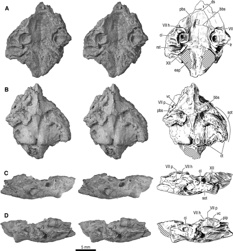
FIGURE 5 Braincase fragment, UA 9684-3 (part of holotype), of Menarana nosymena, gen. et sp. nov., from the Late Cretaceous of Madagascar. Stereophotographs (left and center) and interpretive drawings (right) of A, dorsal; B, ventral; C, left lateral; and D, right lateral views. Abbreviations: bbs, basisphenoid-basioccipital suture; ci, crista interfenestralis; ct, crista tuberalis; ds, dorsum sellae; eap, exoccipital ascending process; lr, lagenar recess; pbs, prootic-basisphenoid suture; pip, inferior process of prootic; rst, recessus scalae tympani; sot, spheno-occipital (basal) tubercle; vc, Vidian canal; VII, facial canal; VII h, foramen for hyomandibular branch of facial nerve; VII p, foramen for palatine branch of facial nerve; and XII, hypoglossal canal.

FIGURE 5 Braincase fragment, UA 9684-3 (part of holotype), of Menarana nosymena, gen. et sp. nov., from the Late Cretaceous of Madagascar. Stereophotographs (left and center) and interpretive drawings (right) of A, dorsal; B, ventral; C, left lateral; and D, right lateral views. Abbreviations: bbs, basisphenoid-basioccipital suture; ci, crista interfenestralis; ct, crista tuberalis; ds, dorsum sellae; eap, exoccipital ascending process; lr, lagenar recess; pbs, prootic-basisphenoid suture; pip, inferior process of prootic; rst, recessus scalae tympani; sot, spheno-occipital (basal) tubercle; vc, Vidian canal; VII, facial canal; VII h, foramen for hyomandibular branch of facial nerve; VII p, foramen for palatine branch of facial nerve; and XII, hypoglossal canal.

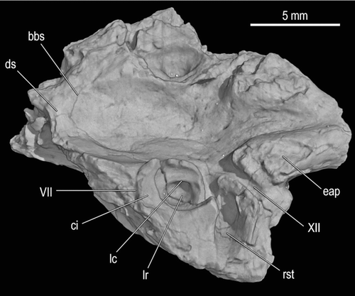
FIGURE 6 Braincase fragment, UA 9684-3 (part of holotype), of Menarana nosymena, gen. et sp. nov., from the Late Cretaceous of Madagascar in dorsolateral view (image obtained from HRXCT dataset). Abbreviations: bbs, basisphenoid-basioccipital suture; ci, crista interfenestralis; ds, dorsum sellae; eap, exoccipital ascending process; lc, lagenar crest; lr, lagenar recess; rst, recessus scalae tympani; VII, facial canal; and XII, hypoglossal canal.

FIGURE 6 Braincase fragment, UA 9684-3 (part of holotype), of Menarana nosymena, gen. et sp. nov., from the Late Cretaceous of Madagascar in dorsolateral view (image obtained from HRXCT dataset). Abbreviations: bbs, basisphenoid-basioccipital suture; ci, crista interfenestralis; ds, dorsum sellae; eap, exoccipital ascending process; lc, lagenar crest; lr, lagenar recess; rst, recessus scalae tympani; VII, facial canal; and XII, hypoglossal canal.

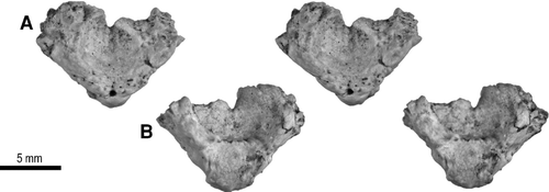
FIGURE 7 Atlas, UA 9684-4 (part of holotype), of Menarana nosymena, gen. et sp. nov., from the Late Cretaceous of Madagascar. Stereophotographs of A, anterior; and B, posterior views.

FIGURE 7 Atlas, UA 9684-4 (part of holotype), of Menarana nosymena, gen. et sp. nov., from the Late Cretaceous of Madagascar. Stereophotographs of A, anterior; and B, posterior views.

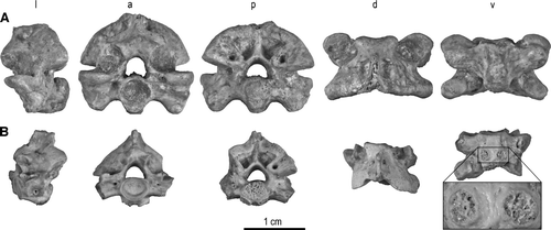
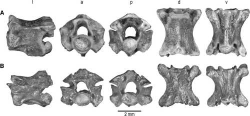
FIGURE 8 Trunk vertebrae of Menarana nosymena, gen. et sp. nov., from the Late Cretaceous of Madagascar in l, lateral; a, anterior; p, posterior; d, dorsal; and v, ventral views. A, anterior trunk vertebra, UA 9687-1 (lateral view reversed); B, mid-trunk vertebra, UA 9684-1 (part of holotype); C, posterior trunk vertebra, UA 9684-2 (part of holotype).

FIGURE 8 Trunk vertebrae of Menarana nosymena, gen. et sp. nov., from the Late Cretaceous of Madagascar in l, lateral; a, anterior; p, posterior; d, dorsal; and v, ventral views. A, anterior trunk vertebra, UA 9687-1 (lateral view reversed); B, mid-trunk vertebra, UA 9684-1 (part of holotype); C, posterior trunk vertebra, UA 9684-2 (part of holotype).

TABLE 2 Measurements of vertebral specimens of Menarana nosymena, gen. et sp. nov. See text for list of abbreviations. ? = vertebral fragment not assigned to region.

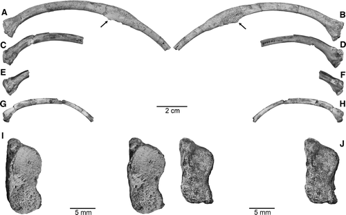
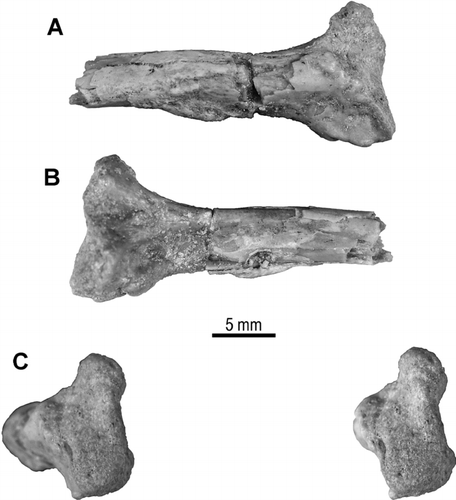
FIGURE 9 Proximal fragment of right rib, UA 9684-5 (part of holotype), of Menarana nosymena, gen. et sp. nov., from the Late Cretaceous of Madagascar in A, anterior; B, posterior, and C, stereophotographic proximal views.

FIGURE 9 Proximal fragment of right rib, UA 9684-5 (part of holotype), of Menarana nosymena, gen. et sp. nov., from the Late Cretaceous of Madagascar in A, anterior; B, posterior, and C, stereophotographic proximal views.

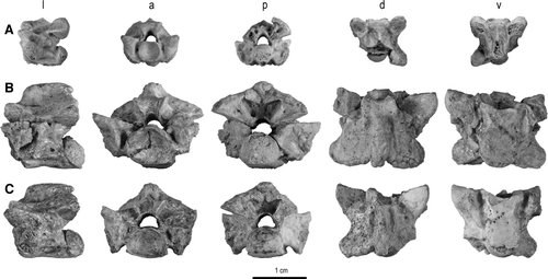
FIGURE 10 Trunk vertebrae of Kelyophis hechti, gen. et sp. nov., from the Late Cretaceous of Madagascar in l, lateral; a, anterior; p, posterior; d, dorsal; and v, ventral views. A, anterior trunk vertebra, FMNH PR 2539 (lateral view reversed); B, mid-trunk vertebra, UA 9682 (holotype).

FIGURE 10 Trunk vertebrae of Kelyophis hechti, gen. et sp. nov., from the Late Cretaceous of Madagascar in l, lateral; a, anterior; p, posterior; d, dorsal; and v, ventral views. A, anterior trunk vertebra, FMNH PR 2539 (lateral view reversed); B, mid-trunk vertebra, UA 9682 (holotype).

TABLE 3 Measurements of vertebral specimens of Kelyophis hechti, gen. et sp. nov. See text for list of abbreviations.

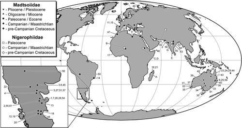
FIGURE 11 Temporal and geographic distribution of madtsoiid (localities indicated by solid symbols, numbers) and nigerophiid (open symbols, letters) snakes. See and for more detailed information.

FIGURE 11 Temporal and geographic distribution of madtsoiid (localities indicated by solid symbols, numbers) and nigerophiid (open symbols, letters) snakes. See Tables 4 and 5 for more detailed information.

TABLE 4 Temporal and geographic distribution of madtsoiid and questionably madtsoiid snakes (arranged alphabetically by genus and species, and then by more uncertain family-level attributions). : = Early; = Middle; = Late.

TABLE 5 Temporal and geographic distribution of nigerophiid and questionably nigerophiid snakes (arranged alphabetically).
Reprints and Corporate Permissions

Please note: Selecting permissions does not provide access to the full text of the article, please see our help page How do I view content?

To request a reprint or corporate permissions for this article, please click on the relevant link below:

Academic Permissions

Please note: Selecting permissions does not provide access to the full text of the article, please see our help page How do I view content?

Obtain permissions instantly via Rightslink by clicking on the button below:

If you are unable to obtain permissions via Rightslink, please complete and submit this Permissions form. For more information, please visit our Permissions help page.