Figures & data
FIGURE 1. Digital representation of the skull of Erlikosaurus andrewsi (IGM 100/111). A, left lateral; B, dorsal; and C, ventral views. Abbreviations: bsp, basisphenoid; exoc, exoccipital; f, frontal; j, jugal; la, lacrimal; mx, maxilla; n, nasal; oc, occipital condyle; pa, parietal; pal, palatine; pmx, premaxilla; po, postorbital; prf, prefrontal; pty, pterygoid; q, quadrate; qj, quadratojugal; soc, supraoccipital; sq, squamosal; vo, vomer.
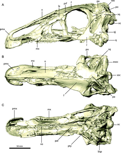
FIGURE 2. Restored skull of Erlikosaurus andrewsi (IGM 100/111). A, left lateral; B, dorsal; and C, ventral views. Abbreviations: bsp, basisphenoid; exoc, exoccipital; f, frontal; j, jugal; la, lacrimal; mx, maxilla; n, nasal; oc, occipital condyle; pa, parietal; pal, palatine; pmx, premaxilla; po, postorbital; prf, prefrontal; pty, pterygoid; q, quadrate; qj, quadratojugal; scr, sclerotic ring; soc, supraoccipital; sq, squamosal; vo, vomer.
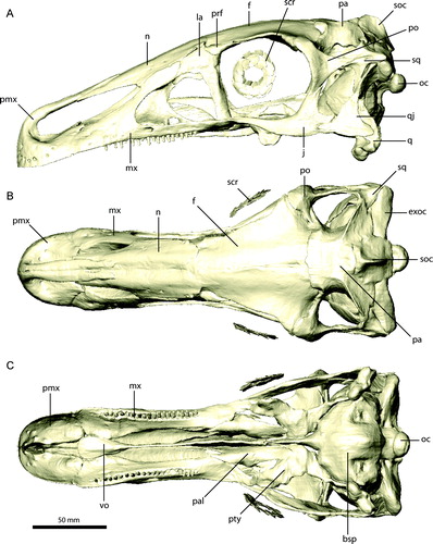
FIGURE 3. Right premaxilla of Erlikosaurus andrewsi (IGM 100/111). A, B, lateral; C, medial; D, dorsal; and E, ventral views. Bone in B rendered transparent to visualize internal neurovascular structures (in yellow). Abbreviations: a.n, nasal articulation; a.pmx, premaxilla articulation; af, ancillary foramina; f1–18, neurovascular foramina; fi, incisive foramen; ml, medial lamina; mp, maxillary process; nc, narial chamber; np, narial process.
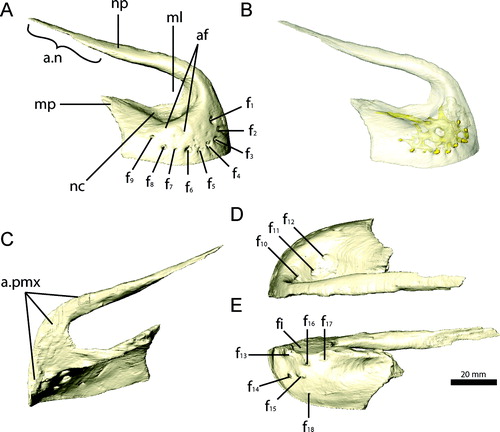
FIGURE 4. Right maxilla of Erlikosaurus andrewsi (IGM 100/111). A, G, lateral; B, F, medial; and C, caudolateral views. 11th maxillary tooth in D, lateral and E, rostral views. Right maxilla fragment of Falcarius utahensis (UMNH VP 14565) in H, medial and I, lateral views. Bone in F–I rendered transparent to visualize teeth (in blue), replacement teeth (in red), and neuropalatine nerve canal (in yellow). Abbreviations: a.pmx, premaxilla articulation; aofo, antorbital fossa; ap, ascending process; cp, caudal process; fo, neurovascular foramina; lnfo, lateral neurovascular foramen; mf, maxillary fenestra; mnfo, medial neurovascular foramen; pnp, pneumatic pockets; ps, palatal shelf.
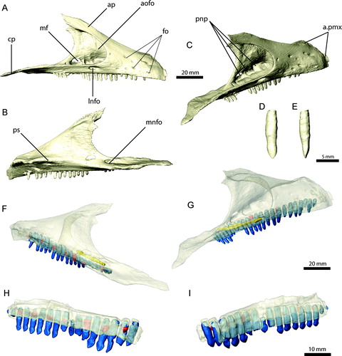
FIGURE 5. Right nasal and left lacrimal of Erlikosaurus andrewsi (IGM 100/111). Right nasal in A, dorsal and B, ventral views. Left lacrimal in C, D, lateral, E, medial, F, rostral, and G, caudal views. Bone in D rendered transparent to visualize internal neurovascular/pneumatic structures (in yellow). Abbreviations: a.f, frontal articulation; a.la, lacrimal articulation; a.mx, maxilla articulation; a.pmx, premaxilla articulation; aofo, antorbital fossa; cp, caudal process; fo, neurovascular foramina; lfo, lacrimal foramen; ra, rostral aperture; rp, rostral process; vp, ventral process.
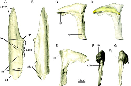
FIGURE 6. Right prefrontal and left jugal of Erlikosaurus andrewsi (IGM 100/111). Right prefrontal in A, lateral, B, dorsal, and C, ventral views. Left jugal in D, lateral and E, medial views. Abbreviations: a.f, frontal articulation; a.la, lacrimal articulation; a.mx, maxilla articulation; a.n, nasal articulation; a.po, postorbital articulation; a.qj, quadratojugal articulation; cp, caudal process; dp, dorsal (postorbital) process; rp, rostral (maxillary) process; vr, ventral ridge.
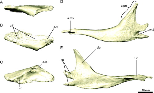
FIGURE 7. Left frontal and parietal of Erlikosaurus andrewsi (IGM 100/111). Left frontal in A, dorsal, B, ventral, C, lateral, and D, medial views. Parietal in E, rostral, F, dorsal, and G, caudal views. Abbreviations: a.f, frontal articulation; a.ls, laterosphenoid articulation; a.n, nasal articulation; a.pa, parietal articulation; a.po, postorbital articulation; a.prf, prefrontal articulation; a.soc, supraoccipital articulation; cc, crista cranii; cer, impression of cerebral hemispheres; lac, lambdoidal crest; ob, impression of olfactory bulbs; orb, orbital cavity; orbm, orbital margin; pop, postorbital process; sqp, squamosal process; stf, supratemporal fenestra; vr, ventral ridge.
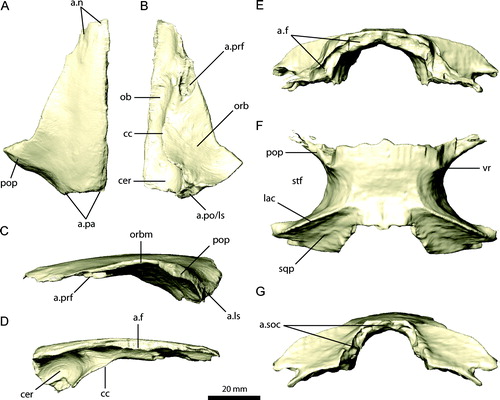
FIGURE 8. Left postorbital, squamosal, and quadratojugal of Erlikosaurus andrewsi (IGM 100/111). Left postorbital in A, lateral, B, medial, and C, caudal views. Left squamosal in D, lateral, E, medial, and F, dorsal views. Left quadratojugal in G, lateral and H, medial views. Abbreviations: a.exoc, exoccipital articulation; a.f, frontal articulation; a.j, jugal articulation; a.ls, laterosphenoid articulation with; a.pa, parietal articulation; a.po, postorbital articulation; a.q, quadrate articulation; a.sq, squamosal articulation; afp, accessory frontal process; dp, dorsal process; fp, frontal process; jp, jugal process; ob, orbit; qp, quadrate process; sqp, squamosal process; stf, supratemporal fenestra.
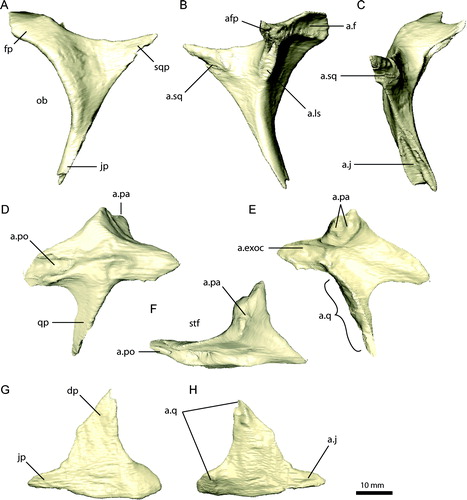
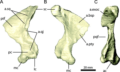
FIGURE 9. Left quadrate of Erlikosaurus andrewsi (IGM 100/111). A, lateral; B, medial; and C, caudal views. Abbreviations: a.bsp, basisphenoid articulation; a.exoc, exoccipital articulation; a.pty, pterygoid articulation; a.qj, quadratojugal articulation; a.sq, squamosal articulation; ac, accessory condyle; lc, lateral condyle; mc, medial condyle; pc, external pneumatic chamber; pqf, paraquadrate foramen; ptf, pterygoid flange; sc, squamosal capitulum.
← FIGURE 10. Braincase of Erlikosaurus andrewsi (IGM 100/111) in A, C, and E, natural articulation and B, D, and F, separated into elements in A, B, caudal, C, D, left lateral, and E, F, rostral views. Abbreviations: boc, basioccipital; bsp, basisphenoid; bsr, basisphenoid recess; ls, laterosphenoid; osp, orbitosphenoid; parp, paroccipital process; pro, prootic; soc, supraoccipital; V1, foramen for ophthalmic branch of the trigeminal nerve; V2, foramen for maxillary branch of the trigeminal nerve; V3, foramen for mandibular branch of the trigeminal nerve; VI, foramen for the abducens nerve; IX–XI, foramina for the glossopharyngal, vagus, and spinal accessory nerves; XII, foramen for the hypoglossal nerve.
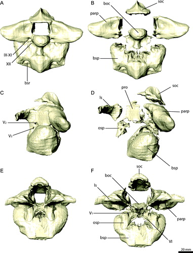
FIGURE 11. Braincase elements of Erlikosaurus andrewsi (IGM 100/111). Supraoccipital in A, dorsal, B, ventral, and C, left lateral views. Right exoccipital/opisthotic in D, rostral, E, G, caudal, and F, dorsal views. Basioccipital in H, caudal, I, dorsal, and J, left lateral views. Bone in G rendered transparent to visualize internal pneumatic structures (in yellow). Abbreviations: a.boc, basioccipital articulation; a.bsp, basisphenoid articulation; a.exoc, exoccipital articulation; a.osp, orbitosphenoid articulation; a.pa, parietal articulation; a.pro, prootic articulation; a.q, quadrate articulation; a.soc, supraoccipital articulation; cp, cerebellar prominence; d, depression; ecf, endocranial floor; nc, nuchal crest; oc, occipital condyle; vr, vertical ridge; stg, stapedial groove; IX–XI, foramina for the glossopharyngal, vagus, and spinal accessory nerves; XII, foramen for the hypoglossal nerve.
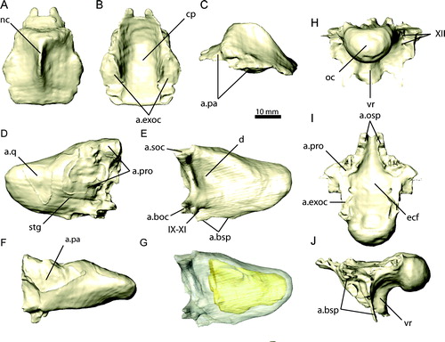
FIGURE 12. Basisphenoid and cultriform process of the parasphenoid of Erlikosaurus andrewsi (IGM 100/111) in A, left lateral, B, ventral, and C, caudal views. Sagittal cross-sections through the braincase of D, Erlikosaurus andrewsi (IGM 100/111) and E, Nothronychus mckinleyi (AZMNH-2117) in left lateral view, revealing pneumatic cavities within the basisphenoid. Abbreviations: bsp, basisphenoid; bsr, basisphenoid recess; cup, cultriform process.
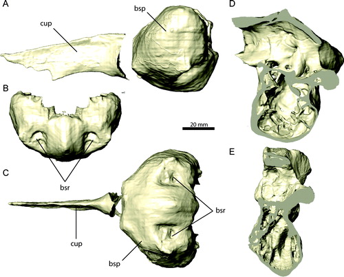
FIGURE 13. Individual braincase elements of Erlikosaurus andrewsi (IGM 100/111). Right prootic (in articulation with exoccipital) in A, lateral and B, medial views. Right laterosphenoid in C, rostral and D, lateral views. Right orbitosphenoid in E, rostral and F, lateral views. Abbreviations: a.bsp, basisphenoid articulation; a.ls, laterosphenoid articulation; a.osp, orbitosphenoid articulation; a.pa, parietal articulation; a.po, postorbital articulation; exoc, exoccipital; fc, fenestra cochleae; ff, floccular fossa; fv, fenestra vestibuli; pop, postorbital process; pro, prootic; rr, rostral horizontal ridge; V1, foramen for ophthalmic branch of the trigeminal nerve; V2, foramen for maxillary branch of the trigeminal nerve; V3, foramen for mandibular branch of the trigeminal nerve; VI, foramen for the abducens nerve; VII, foramen for the facial nerve; VIII, foramen for the vestibulocochlear nerve.
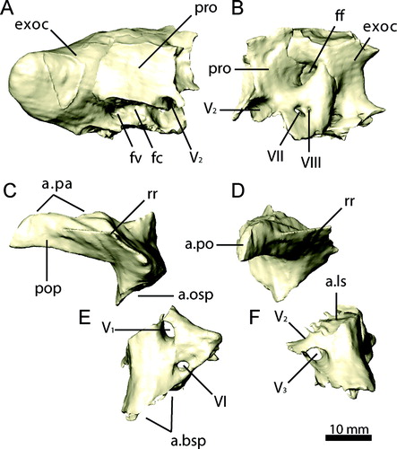
FIGURE 14. Palatal complex of Erlikosaurus andrewsi (IGM 100/111) in A, dorsal, B, left lateral, and C, ventral views. Abbreviations: cp, cultriform process; ect, ectopterygoid; pal, palatine; pty, pterygoid; spf, subsidiary palatal fenestra; vo, vomer.
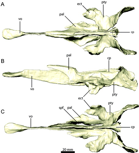
FIGURE 15. Palatal elements of Erlikosaurus andrewsi (IGM 100/111). Vomer in A, left lateral and B, dorsal views. Right palatine in C, dorsal and D, lateral views. Right pterygoid and ectopterygoid in E, dorsal and F, lateral views. Abbreviations: a.bsp, basisphenoid articulation; a.cp/vpar, cultriform process and vomeropalatine ramus of the pterygoid articulation; a.ect, articulation with ectopterygoid; a.j, jugal articulation; a.max, maxilla articulation; a.pmx/mx, premaxilla/maxilla articulation; a.pty, pterygoid articulation; a.q, quadrate articulation; a.vpal, vomeropterygoid ramus of the palatine articulation; ect, ectopterygoid; ectr, ectropterygoid ramus; papr, palatine pneumatic recess; ppp, pterygoid process of palatine; qr, quadrate ramus; spf, subsidiary palatal fenestra; tf, transverse flange; vpal, vomeropterygoid ramus; vpar, vomeropalatine ramus.
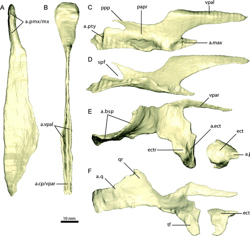
FIGURE 16. Sclerotic elements of Erlikosaurus andrewsi (IGM 100/111). A, articulated sclerotic plates as preserved (six of seven preserved elements shown). Isolated, single sclerotic plate in B, lateral and C, medial views. D, fully reconstructed sclerotic ring.
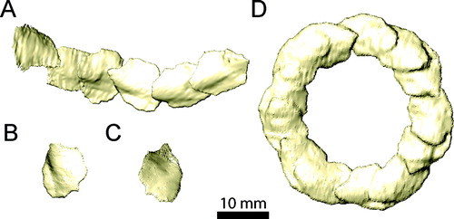
FIGURE 17. Left mandible of Erlikosaurus andrewsi (IGM 100/111) in A, lateral, B, medial, and C, dorsal views. Abbreviations: ang, angular; ar, articular; d, dentary; emf, external mandibular fenestra; maf, mandibular fossa; pra, prearticular; sp, splenial; sur, surangular.
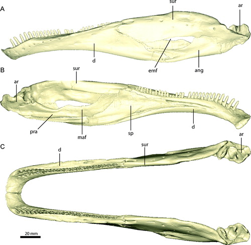
FIGURE 18. Left dentary of Erlikosaurus andrewsi (IGM 100/111) in A, C, lateral, and B, D, medial views. Right dentaries of Falcarius utahensis in medial view: E, UMNH VP 14527, F, UMNH VP 14529, G, UMNH VP 14528. Bone in C–G rendered transparent to visualise teeth (in blue), replacement teeth (in red), and neurovascular structures (in yellow). Abbreviations: a.pra, prearticular articulation; a.sur, surangular articulation; fo, neurovascular foramina; lsh, lateral shelf; mf, Meckelian fossa; sf, symphyseal facet.
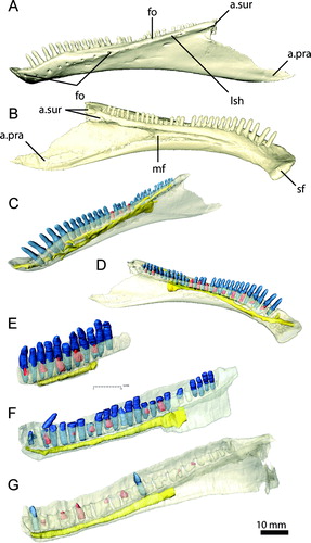
FIGURE 19. Mandibular elements of Erlikosaurus andrewsi (IGM 100/111). Left splenial in A, medial and B, lateral views. Left surangular in C, D, lateral, E, dorsal, and F, medial views. Left angular in G, lateral and H, medial views. Left prearticular in I, medial and J, lateral views. Left articular in K, lateral, L, medial, and M, dorsal views. Bone in F rendered transparent to visualize internal neurovascular structures (in yellow). Abbreviations: a.ang, angular articulation; a.art, articular articulation; a.d, dentary articulation; a.pra, prearticular articulation; a.sp, splenial articulation; a.sur, surangular articulation; cgb, caudal glenoid buttress; clf, caudolateral foramen; cp, caudal process; de, elongated depression; dfo, dorsal foramen; dr, dorsal ridge; emf, external mandibular fenestra; fmf, floor of mandibular fossa; fo, foramen for the chorda tympani; g, groove; igr, interglenoid ridge; maf, mandibular adductor fossa; mf, medial foramen; mr, medial ridge; nif, noninvasive foramina; rap, retroarticular process; rf, rostral foramina; rgb, rostral glenoid buttress tp, transverse process; vfo, ventral foramen; vr, ventral ridge.
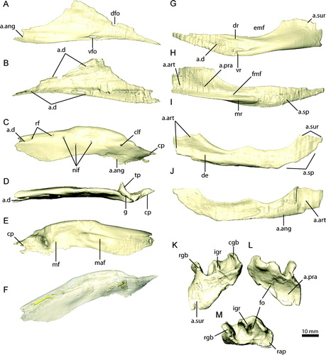
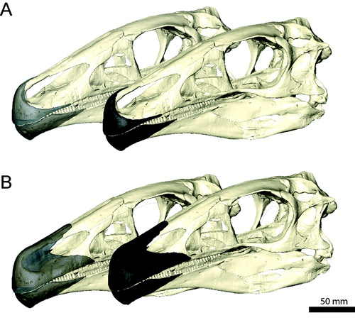
FIGURE 20. Reconstructed rhamphothecae of Erlikosaurus andrewsi with A, small and B, extensive keratinous sheath (rendered transparent in the back to reveal underlying bone).