2,525
Views
15
CrossRef citations to date
0
Altmetric
Articles

A reassessment of the postcanine dentition and systematics of the tritylodontid Stereognathus (Cynodontia, Tritylodontidae, Mammaliamorpha), from the Middle Jurassic of the United Kingdom

ORCID Icon, , ORCID Icon, &
Article: e1351448 | Received 06 Apr 2017, Accepted 10 Jun 2017, Published online: 12 Sep 2017

Figures & data

TABLE 1. Measurements of U.K. Stereognathus material.

FIGURE 1. Postcanine cusp terminology for Stereognathus used in this paper (modified from Watabe et al., Citation2007). The PIA (posterior interlocking area) and AIA (anterior interlocking area) are present on both teeth, but the PIA is not visible in occlusal view on upper postcanines and the AIA is not visible in occlusal view on lower postcanines, because they are located on the underside of the tooth.
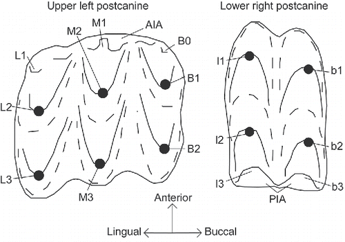
FIGURE 1. Postcanine cusp terminology for Stereognathus used in this paper (modified from Watabe et al., Citation2007). The PIA (posterior interlocking area) and AIA (anterior interlocking area) are present on both teeth, but the PIA is not visible in occlusal view on upper postcanines and the AIA is not visible in occlusal view on lower postcanines, because they are located on the underside of the tooth.

FIGURE 2. Stereognathus ooliticus, BGS GSM113834, holotype. A1, occlusal view; A2, occlusal view digital reconstruction, with teeth segmented from jaw; B1, buccal view; B2, buccal view digital reconstruction, with teeth segmented from jaw; C1, lingual view; C2, lingual view digital reconstruction, with teeth segmented from jaw; D, dorsal view of maxilla. Anterior direction indicated by longer black arrow, lingual by shorter arrow.
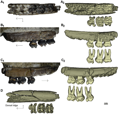
FIGURE 2. Stereognathus ooliticus, BGS GSM113834, holotype. A1, occlusal view; A2, occlusal view digital reconstruction, with teeth segmented from jaw; B1, buccal view; B2, buccal view digital reconstruction, with teeth segmented from jaw; C1, lingual view; C2, lingual view digital reconstruction, with teeth segmented from jaw; D, dorsal view of maxilla. Anterior direction indicated by longer black arrow, lingual by shorter arrow.

FIGURE 3. Stereognathus ooliticus, BGS GSM113834, holotype, postcanines only. Digitally reconstructed from microCT scans and segmented from the jaw. A, occlusal view of anterior-most postcanine; B, occlusal view of middle postcanine; C, occlusal view of posterior-most postcanine; D, original drawing by Owen (Citation1857); E, occlusal view digital reconstruction; F, dorsal view digital reconstruction; G, anterolingual view digital reconstruction; H1, anterior view of anterior-most postcanine digital reconstruction; H2, posterior view of anterior-most postcanine digital reconstruction; I1, anterior view of middle postcanine digital reconstruction; I2, posterior view of middle postcanine digital reconstruction; J1, anterior view of posterior-most postcanine digital reconstruction; J2, posterior view of posterior-most postcanine digital reconstruction. Anterior direction indicated by longer black arrow, lingual by shorter arrow.
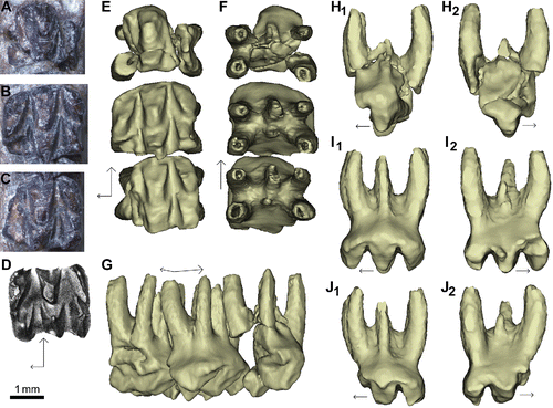
FIGURE 3. Stereognathus ooliticus, BGS GSM113834, holotype, postcanines only. Digitally reconstructed from microCT scans and segmented from the jaw. A, occlusal view of anterior-most postcanine; B, occlusal view of middle postcanine; C, occlusal view of posterior-most postcanine; D, original drawing by Owen (Citation1857); E, occlusal view digital reconstruction; F, dorsal view digital reconstruction; G, anterolingual view digital reconstruction; H1, anterior view of anterior-most postcanine digital reconstruction; H2, posterior view of anterior-most postcanine digital reconstruction; I1, anterior view of middle postcanine digital reconstruction; I2, posterior view of middle postcanine digital reconstruction; J1, anterior view of posterior-most postcanine digital reconstruction; J2, posterior view of posterior-most postcanine digital reconstruction. Anterior direction indicated by longer black arrow, lingual by shorter arrow.
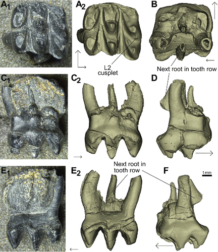
FIGURE 4. Stereognathus hebridicus, BRSUG 20572, holotype. A1, occlusal view; A2, occlusal view digital reconstruction; B, dorsal view digital reconstruction; C1, anterior view; C2, anterior view digital reconstruction; D, buccal view digital reconstruction; E1, posterior view; E2, posterior view digital reconstruction; F, lingual view digital reconstruction. Anterior direction indicated by longer black arrow, lingual by shorter arrow.
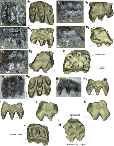
FIGURE 4. Stereognathus hebridicus, BRSUG 20572, holotype. A1, occlusal view; A2, occlusal view digital reconstruction; B, dorsal view digital reconstruction; C1, anterior view; C2, anterior view digital reconstruction; D, buccal view digital reconstruction; E1, posterior view; E2, posterior view digital reconstruction; F, lingual view digital reconstruction. Anterior direction indicated by longer black arrow, lingual by shorter arrow.
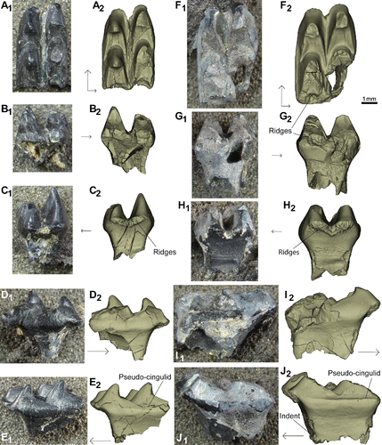
FIGURE 5. Stereognathus hebridicus, BRSUG 20573, paratype, upper postcanines, and new specimen NMS G.2017.17.2, both reconstructed digitally from micro CT scans. AF, BRSUG 20573: A1, occlusal view; A2, occlusal view digital reconstruction; B1, anterior view; B2, anterior view digital reconstruction; C1, posterior view; C2, posterior view digital reconstruction; D1, lingual view; D2, lingual view digital reconstruction; E1, buccal view; E2, buccal view digital reconstruction; F, dorsal view. GM, NMS G.2017.17.2: G1, occlusal view; G2, occlusal view digital reconstruction; H1, anterior view; H2, anterior view digital reconstruction; I, posterior view digital reconstruction; J, lingual view digital reconstruction; K, buccal view digital reconstruction; L, dorsal view digital reconstruction; M, ventrolingual view digital reconstruction. Anterior direction indicated by longer black arrow, lingual by shorter arrow.
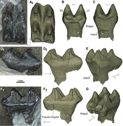
FIGURE 5. Stereognathus hebridicus, BRSUG 20573, paratype, upper postcanines, and new specimen NMS G.2017.17.2, both reconstructed digitally from micro CT scans. A–F, BRSUG 20573: A1, occlusal view; A2, occlusal view digital reconstruction; B1, anterior view; B2, anterior view digital reconstruction; C1, posterior view; C2, posterior view digital reconstruction; D1, lingual view; D2, lingual view digital reconstruction; E1, buccal view; E2, buccal view digital reconstruction; F, dorsal view. G–M, NMS G.2017.17.2: G1, occlusal view; G2, occlusal view digital reconstruction; H1, anterior view; H2, anterior view digital reconstruction; I, posterior view digital reconstruction; J, lingual view digital reconstruction; K, buccal view digital reconstruction; L, dorsal view digital reconstruction; M, ventrolingual view digital reconstruction. Anterior direction indicated by longer black arrow, lingual by shorter arrow.

FIGURE 6. Stereognathus hebridicus, BRSUG 20574 and BRSUG 20575, paratypes, lower postcanines. AE, BRSUG 20574: A1, occlusal view; A2, occlusal view digital reconstruction; B1, posterior view; B2, posterior view digital reconstruction; C1, anterior view; C2, anterior view digital reconstruction; D1, lingual view; D2, lingual view digital reconstruction; E1, buccal view; E2, buccal view digital reconstruction. FJ, BRSUG 20575: F1, occlusal view; F2, occlusal view digital reconstruction; G1, posterior view; G2, posterior view digital reconstruction; H1, anterior view; H2, anterior view digital reconstruction; I1, lingual view; I2, lingual view digital reconstruction; J1, buccal view; J2, buccal view digital reconstruction. Anterior direction indicated by longer black arrow, lingual by shorter arrow.

FIGURE 6. Stereognathus hebridicus, BRSUG 20574 and BRSUG 20575, paratypes, lower postcanines. A–E, BRSUG 20574: A1, occlusal view; A2, occlusal view digital reconstruction; B1, posterior view; B2, posterior view digital reconstruction; C1, anterior view; C2, anterior view digital reconstruction; D1, lingual view; D2, lingual view digital reconstruction; E1, buccal view; E2, buccal view digital reconstruction. F–J, BRSUG 20575: F1, occlusal view; F2, occlusal view digital reconstruction; G1, posterior view; G2, posterior view digital reconstruction; H1, anterior view; H2, anterior view digital reconstruction; I1, lingual view; I2, lingual view digital reconstruction; J1, buccal view; J2, buccal view digital reconstruction. Anterior direction indicated by longer black arrow, lingual by shorter arrow.

FIGURE 7. New specimen NMS G.1992.47.120, a lower postcanine, reconstructed digitally from micro CT scans. A1, occlusal view; A2, occlusal view digital reconstruction; B, posterior view digital reconstruction; C, anterior view digital reconstruction; D1, lingual view; D2, lingual view digital reconstruction; E, anterolingual view digital reconstruction; F1, buccal view; F2, buccal view digital reconstruction; G, posterobuccal view digital reconstruction. Anterior direction indicated by longer black arrow, lingual by shorter arrow.

FIGURE 7. New specimen NMS G.1992.47.120, a lower postcanine, reconstructed digitally from micro CT scans. A1, occlusal view; A2, occlusal view digital reconstruction; B, posterior view digital reconstruction; C, anterior view digital reconstruction; D1, lingual view; D2, lingual view digital reconstruction; E, anterolingual view digital reconstruction; F1, buccal view; F2, buccal view digital reconstruction; G, posterobuccal view digital reconstruction. Anterior direction indicated by longer black arrow, lingual by shorter arrow.

FIGURE 8. Scatterplots of postcanine measurements of Stereognathus. A, upper postcanines; B, lower postcanines. Key for B, as in A. Solid symbols denote complete specimens; open symbols denote incomplete specimens (orange square = S. ooliticus; blue diamond = S. hebridicus). Measurements in .
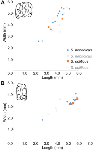
FIGURE 8. Scatterplots of postcanine measurements of Stereognathus. A, upper postcanines; B, lower postcanines. Key for B, as in A. Solid symbols denote complete specimens; open symbols denote incomplete specimens (orange square = S. ooliticus; blue diamond = S. hebridicus). Measurements in Table 1.

FIGURE 9. Distributions of dimensions of Stereognathus postcanine specimens. A, upper postcanine length; B, upper postcanine width; C, lower postcanine length; D, lower postcanine width. Orange striped bars = S. ooliticus; blue bars = S. hebridicus.
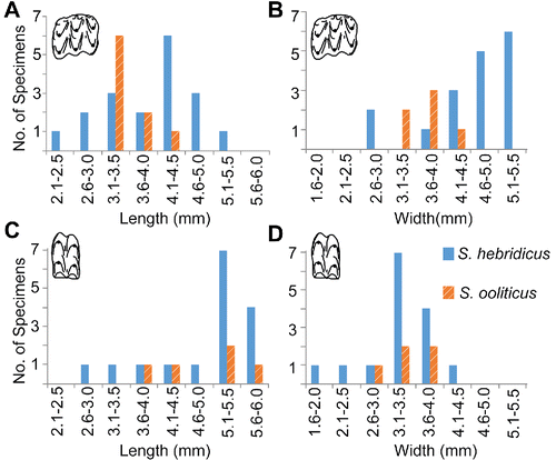
FIGURE 9. Distributions of dimensions of Stereognathus postcanine specimens. A, upper postcanine length; B, upper postcanine width; C, lower postcanine length; D, lower postcanine width. Orange striped bars = S. ooliticus; blue bars = S. hebridicus.

TABLE 2. The data set used for analysis, including estimated measurements. Measurements in mm.

FIGURE 10. Trees generated by our phylogenetic analysis of tritylodontid taxa, using updated character codings for Stereognathus. A, strict consensus of the five parsimonious trees of 71 steps; B, the agreement subtree of 10 taxa.
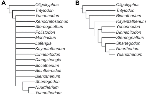
FIGURE 10. Trees generated by our phylogenetic analysis of tritylodontid taxa, using updated character codings for Stereognathus. A, strict consensus of the five parsimonious trees of 71 steps; B, the agreement subtree of 10 taxa.

APPENDIX 2. Character matrix used for phylogenetic analysis. Polymorphisms are as follows: A (0, 2); B (2, 6); C (1, 0); D (2, 3)