2,351
Views
2
CrossRef citations to date
0
Altmetric
Articles

A new genus and species of extinct ground shark, †Diprosopovenator hilperti, gen. et sp. nov. (Carcharhiniformes, †Pseudoscyliorhinidae, fam. nov.), from the Upper Cretaceous of Germany

ORCID Icon, & ORCID Icon
Article: e1593185 | Received 27 Nov 2018, Accepted 26 Feb 2019, Published online: 03 May 2019

Figures & data

FIGURE 1. Geographic and stratigraphic information. A, B, maps of geographic location. C, Cenomanian to Turonian lithostratigraphic subdivision in Lower Saxony (compiled after Niebuhr et al., Citation2007, and Ogg et al., Citation2016). D, lithological section of the upper Cenomanian–lower Turonian transition formerly exposed in the HPCF II quarry of Hanover-Misburg (after Ernst et al., Citation1984; Maisch and Lehmann, Citation2000), with stratigraphic position of †Diprosopovenator hilperti, gen. et sp. nov., indicated by the star.

FIGURE 1. Geographic and stratigraphic information. A, B, maps of geographic location. C, Cenomanian to Turonian lithostratigraphic subdivision in Lower Saxony (compiled after Niebuhr et al., Citation2007, and Ogg et al., Citation2016). D, lithological section of the upper Cenomanian–lower Turonian transition formerly exposed in the HPCF II quarry of Hanover-Misburg (after Ernst et al., Citation1984; Maisch and Lehmann, Citation2000), with stratigraphic position of †Diprosopovenator hilperti, gen. et sp. nov., indicated by the star.

FIGURE 2. Diprosopovenator hilperti, gen. et sp. nov., RE A 4872/1, holotype. A, interpretative composite drawing combining RE A 4872/1 (dark gray) and RE A 4872/2 (light gray; see ). BE, RE A 4872/1. B, under normal light; C, close-up view of placoid scales; D, interpretative drawing; E, under ultraviolet light. Abbreviations: ao.w, antorbital wall; d, dermis; hym, hyomandibula; l, left (in parentheses, e.g., ‘M.c(l)’); M.c, Meckel’s cartilage; nc, neurocranium; ot.cap, otic capsule; pl.sc; placoid scales; po.pr, postorbital process; po.w, postorbital wall; pq, palatoquadrate; r, right (in parentheses, e.g., ‘M.c(r)’); so.cr, supraorbital crest; spt.r, sphenopterotic ridge; t, teeth; v.c, vertebral centra.

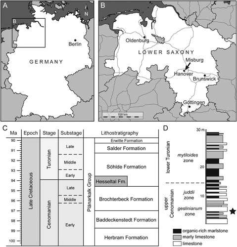
FIGURE 2. †Diprosopovenator hilperti, gen. et sp. nov., RE A 4872/1, holotype. A, interpretative composite drawing combining RE A 4872/1 (dark gray) and RE A 4872/2 (light gray; see Fig. 3). B–E, RE A 4872/1. B, under normal light; C, close-up view of placoid scales; D, interpretative drawing; E, under ultraviolet light. Abbreviations: ao.w, antorbital wall; d, dermis; hym, hyomandibula; l, left (in parentheses, e.g., ‘M.c(l)’); M.c, Meckel’s cartilage; nc, neurocranium; ot.cap, otic capsule; pl.sc; placoid scales; po.pr, postorbital process; po.w, postorbital wall; pq, palatoquadrate; r, right (in parentheses, e.g., ‘M.c(r)’); so.cr, supraorbital crest; spt.r, sphenopterotic ridge; t, teeth; v.c, vertebral centra.

FIGURE 3. Diprosopovenator hilperti, gen. et sp. nov., RE A 4872/2, holotype. A, under normal light; B, close-up view of vertebral column; C, under ultraviolet light. Abbreviations: l, left (in parentheses); M.c, Meckel’s cartilage; r, right (in parentheses).

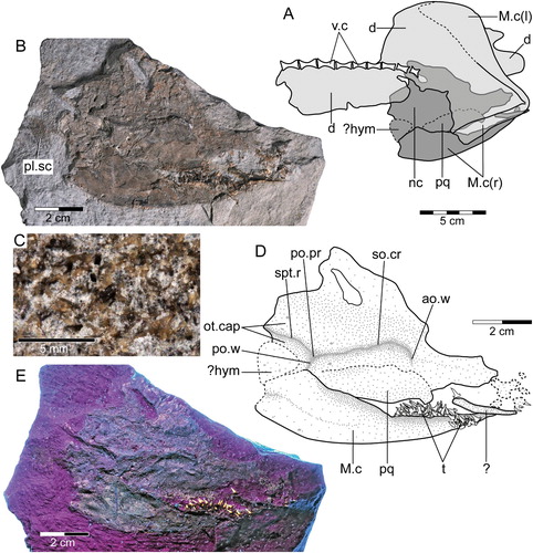
FIGURE 3. †Diprosopovenator hilperti, gen. et sp. nov., RE A 4872/2, holotype. A, under normal light; B, close-up view of vertebral column; C, under ultraviolet light. Abbreviations: l, left (in parentheses); M.c, Meckel’s cartilage; r, right (in parentheses).

FIGURE 4. Diprosopovenator hilperti, gen. et sp. nov., RE A 4872/1 and RE A 4872/2, holotype. Close-up views of dentitions preserved in A, RE A 4872/1 (anterior to right) and B, RE A 4872/2 (anterior to top). Abbreviations: d.gr, dental groove; l, left (in parentheses, e.g., ‘M.c(l)’); M.c, Meckel’s cartilage; pq, palatoquadrate; r, right (in parentheses, e.g., ‘M.c(r)’).

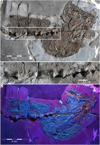
FIGURE 4. †Diprosopovenator hilperti, gen. et sp. nov., RE A 4872/1 and RE A 4872/2, holotype. Close-up views of dentitions preserved in A, RE A 4872/1 (anterior to right) and B, RE A 4872/2 (anterior to top). Abbreviations: d.gr, dental groove; l, left (in parentheses, e.g., ‘M.c(l)’); M.c, Meckel’s cartilage; pq, palatoquadrate; r, right (in parentheses, e.g., ‘M.c(r)’).

FIGURE 5. Diprosopovenator hilperti, gen. et sp. nov., RE A 4872/1 and RE A 4872/2, holotype. Close-up views of selected teeth preserved in AD, RE A 4872/1 and EJ, RE A 4872/2 under normal light. A, two anterolateral teeth in basal and labial views, respectively. B, lateral teeth in labial views. C, posterior tooth in labial view. D, posterior tooth with broken main cusp in labial view. E, anterior tooth in labial view. F, anterolateral tooth in oblique view. G, lateral tooth in basal view. H, lateral tooth in oblique view. I, posterolateral tooth in lingual view. J, lateral tooth in lingual view with broken main cusp. KO, SEM pictures of lateral tooth extracted from RE A 4872/2 in K, labial, L, occlusal, M, labial, N, mesial, and, O, distal views. Pulp cavities marked by arrows.

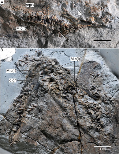
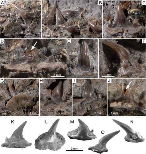
FIGURE 5. †Diprosopovenator hilperti, gen. et sp. nov., RE A 4872/1 and RE A 4872/2, holotype. Close-up views of selected teeth preserved in A–D, RE A 4872/1 and E–J, RE A 4872/2 under normal light. A, two anterolateral teeth in basal and labial views, respectively. B, lateral teeth in labial views. C, posterior tooth in labial view. D, posterior tooth with broken main cusp in labial view. E, anterior tooth in labial view. F, anterolateral tooth in oblique view. G, lateral tooth in basal view. H, lateral tooth in oblique view. I, posterolateral tooth in lingual view. J, lateral tooth in lingual view with broken main cusp. K–O, SEM pictures of lateral tooth extracted from RE A 4872/2 in K, labial, L, occlusal, M, labial, N, mesial, and, O, distal views. Pulp cavities marked by arrows.