Figures & data
FIGURE 1. Holotype of Danielsraptor phorusrhacoides, gen. et sp. nov. from the lower Eocene London Clay of Walton-on-the-Naze, Essex, U.K. (NMS.Z.2021.40.12), skull elements. A, B, upper beak in A, lateral and B, ventral view. C, dorsal nasal bar in ventral view. D, jugal bars. E, F, dorsal portion of neurocranium in dorsal (E) and ventral (F) view. G, H, mandible in dorsal (G) and lateral (H) view; the arrow denotes an enlarged view of the caudal end. I–L, left quadrate in caudal (I), medial (J), lateral (K), and ventral (L) view. M, skull of Masillaraptor parvunguis from the Messel fossil site in Germany (IRSNB Av 83). N, O, left os lacrimale in dorsal (N) and lateral (O) view. P, two unidentified ossicles, which may represent the caudal portions of the pterygoids. Abbreviations: cdl, condylus lateralis; cdm, condylus medialis; cdp, condylus pterygoideus; cpo, capitulum oticum; cps, capitulum squamosum; ctq, cotyla quadratojugalis; ctl, cotyla lateralis; dcp, descending process of lacrimal; fpn, foramen pneumaticum; pmd, processus medialis; ppo, processus postorbitalis; prj, dorsal projection; pso, processus supraorbitalis; tgr, tomial groove. Scale bars equal 10 mm; same scale for A–H.
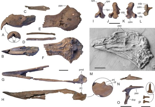
FIGURE 2. Comparison of skull elements of the Masillaraptoridae, crown group Falconiformes, and the Cariamiformes. A, digitally assembled skull bones and mandible of A, Danielsraptor phorusrhacoides (holotype, NMS.Z.2021.40.12). B, skull of Caracara plancus (Falconiformes; SMF 3462). C, skull of Psilopterus lemoinei (Cariamiformes, Phorusrhacidae; AMNH 9257, the dotted line indicates the shape of the missing tip of the beak). D–L, left quadrate of D–F, D. phorusrhacoides (holotype), G–I, C. plancus (SMF 6441), and J–L, Cariama cristata (Cariamiformes; SMF 1862; right quadrate, mirrored) in caudal (D, G, J), medial (E, H, K), and lateral (F, I, L) view. M–O, caudal end of mandible (lateral view) of M, D. phorusrhacoides (holotype), N, Micrastur ruficollis (Falconiformes; SMF 9812), and O, C. cristata (SMF 1862). Abbreviations: cdl, condylus lateralis; cdm, condylus medialis; cdp, condylus pterygoideus; cpo, capitulum oticum; cps, capitulum squamosum; iic, incisura intercapitularis; orb, processus orbitalis; prj, dorsal projection; tgr, tomial groove. Scale bars equal 10 mm.
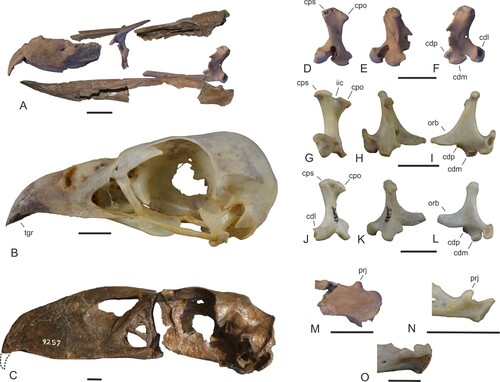
FIGURE 3. Holotype of Danielsraptor phorusrhacoides, gen. et sp. nov. from the lower Eocene London Clay of Walton-on-the-Naze, Essex, U.K. (NMS.Z.2021.40.12), vertebrae and elements of the pectoral girdle and wing. A–D, four thoracic vertebrae in dorsal and lateral view. E, a caudal vertebra with a long processus ventralis in cranial, ventral, and lateral view. F, five further caudal vertebrae in different views. G, pygostyle in lateral view. H, I, left coracoid in dorsal (H) and ventral (I) view. J–L, right coracoid in ventral (J), dorsal (K), and medial (L) view. M, N, right scapula in medial (M) and lateral (N) view. O, extremitas sternalis of furcula. P, left extremitas omalis of furcula. Q, R, cranialmost portion of sternum in lateral (Q) and ventral (R) view. S, proximal portion of left humerus in cranial view. T, U, distal end of left (T) and right (U) humerus in cranial view. V, distal end of left ulna in ventral view. Abbreviations: cdd, condylus dorsalis; cdv, condylus ventralis; csc, cotyla scapularis; fac, facies articularis clavicularis; fns, foramen nervi supracoracoidei; fos, fossa in sulcus supracoracoideus; pcv, processus ventralis; sac, sulcus articularis coracoideus; spe, spina externa. Scale bar equals 10 mm.
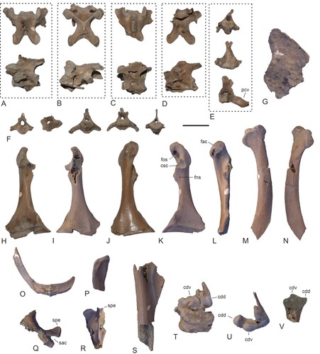
FIGURE 4. Danielsraptor phorusrhacoides, gen. et sp. nov., bones of the tentatively referred specimen NMS.Z.2021.40.13 in comparison to the holotype and Masillaraptor cf. parvunguis. A, three thoracic vertebrae of NMS.Z.2021.40.13 in lateral and—regarding a fragmentary specimen—dorsal view. B, seven caudal vertebrae of NMS.Z.2021.40.13 in different views. C, furcula of NMS.Z.2021.40.13. D–F, pygostyle of D, the holotype of D. phorusrhacoides (NMS.Z.2021.40.12), E, the tentatively referred specimen NMS.Z.2021.40.13, and F, M. cf. parvunguis (NMS.Z.2021.40.14); the dotted lines indicate the reconstructed hypothetical outlines of the bones. G–I, coracoid (dorsal view) of G, the holotype of D. phorusrhacoides, H, the tentatively referred specimen NMS.Z.2021.40.13, and I, M. cf. parvunguis (NMS.Z.2021.40.14). J, K, right scapula (lateral view) of J, the holotype of D. phorusrhacoides and K, the tentatively referred specimen NMS.Z.2021.40.13. L–N, sternum fragments (ventral view) of L, the holotype of D. phorusrhacoides and M, N, the tentatively referred specimen NMS.Z.2021.40.13 (M: corpus in ventral view, N: caudolateral portion of left side); the arrows denote details of the spina externa. O, P, distal end of the humerus (cranial view) of O, M. cf. parvunguis (left side; NMS.Z.2021.40.14) and P, the holotype of D. phorusrhacoides (right side). Abbreviations: acr, acromion; fah, facies articularis humeralis; flx, processus flexorius; icl, incisura lateralis; icm, incisura medialis; mpr, medial projection; pcc, processus costales; pcv, processus ventralis; per, perforation in caudoventral portion of pygostyle; ppc, processus procoracoideus; spe, spina externa; spr, sternal projection on tip of processus procoracoideus; tbi, trabecula intermedia; tbl, trabecula lateralis. Scale bars equal 10 cm.
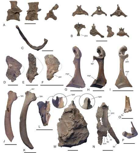
FIGURE 5. Holotype of Danielsraptor phorusrhacoides, gen. et sp. nov. from the lower Eocene London Clay of Walton-on-the-Naze, Essex, U.K. (NMS.Z.2021.40.12), pelvis and hindlimb elements. A, B, pelvis (dorsal view) and left femur in a block of matrix in dorsal (A) and lateral (B) view; C, D, right femur in cranial (C) and caudal (D) view. E, left tibiotarsus lacking distal end. F–H, left tarsometatarsus in dorsolateral (F), dorsal (G), and plantar (H) view. I, J, right tarsometatarsus in dorsal (I) and medial (J) view. K, distal end of right tarsometatarsus in distal view. L, proximal end of right tarsometatarsus in proximal view. M, os metatarsale I and pedal phalanges of the left foot. N, second phalanges of third toe (left and right). O, os metatarsale I and pedal phalanges of the extant Caracara plancus (Falconiformes, SMF 4092). P, os metatarsale I and pedal phalanges of the extant Cariama cristata (Cariamiformes; SMF 2462, the arrows delimit the borders between articulated phalanges of the fourth toe). The identity of the phalanges in M, O, and P is indicated by Roman (toes) and Arabic (phalanges) numerals. Abbreviations: ais, ala ischii; att, antitrochanter; cst, constriction; ctr, crista trochanteris; fac, foramen acetabuli; fii, foramen ilioischiadicum; flg, plantarly directed flange of trochlea metatarsi IV; fob, foramen obturatum; lfe, left femur; lip, lip-like projection formed by cotyla; mtI, os metatarsale I; pub, pubis; sul, hypotarsal sulcus for tendon of musculus flexor digitorum longus. Scale bars equal 10 mm (same scale for A–J).
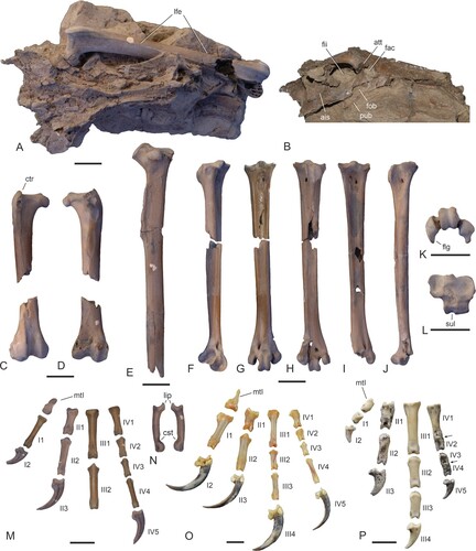
FIGURE 6. Leg bones of Masillaraptor cf. parvunguis from the lower Eocene London Clay of Walton-on-the-Naze, Essex, U.K. (A–I) compared with other fossils of Masillaraptor (J, K), as well as London Clay specimens of Coturnipes (L, M), Danielsraptor, gen. nov. (N, O), and Stintonornis (P, Q). A, Masillaraptor cf. parvunguis, proximal end of left femur in cranial view (NMS.Z.2021.40.14). B, M. cf. parvunguis, distal end of right femur in caudal view (NMS.Z.2021.40.14). C, M. cf. parvunguis, distal end of left tibiotarsus in cranial view (NMS.Z.2021.40.14). D, M. cf. parvunguis, distal end of right tibiotarsus in cranial view (NMS.Z.2021.40.14). E, F, M. cf. parvunguis, left tarsometatarsus in dorsal (E) and plantar (F) view (NMS.Z.2021.40.14). G–I, M. cf. parvunguis, right tarsometatarsus in dorsal (G), plantar (H), and distal (I) view (NMS.Z.2021.40.14). J, left tarsometatarsus of Masillaraptor sp. from Messel in plantar view (SMF-ME 1068). K, left tarsometatarsus of Masillaraptor sp. from the middle Eocene of the Geisel Valley in plantar view (GMH NW XIV). L, M, distal end of the left tarsometatarsus of Coturnipes cooperi from the London Clay of the Isle of Sheppey in L, dorsal and M, plantar view (holotype, NHMUK A 3706). N, O, right tarsometatarsus of Danielsraptor phorusrhacoides, gen. et sp. nov. (NMS.Z.2021.40.12) in N, plantar and O, dorsal view. P, Q, distal end of the right tarsometatarsus of Stintonornis mitchelli from the London Clay of the Isle of Sheppey in P, plantar and Q, dorsal view (holotype, NHMUK A 5284). R, pedal phalanges of Masillaraptor cf. parvunguis (NMS.Z.2021.40.14); the identity of some phalanges is indicated by Roman (toes) and Arabic (phalanges) numerals. For the tarsometatarsi, the trochleae are numbered. Abbreviations: fvd, foramen vasculare distale; mtI, fossa metatarsi I; pst, pons supratendineus. Scale bars equal 5 mm.
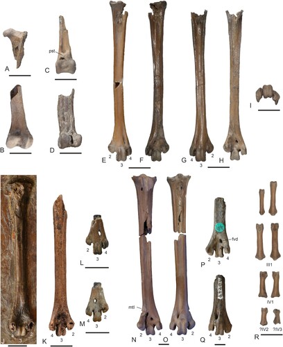
FIGURE 7. Vertebrae as well as pectoral girdle and wing elements of Masillaraptor cf. parvunguis from the lower Eocene London Clay of Walton-on-the-Naze, Essex, U.K. A, three cervical vertebrae in different views (NMS.Z.2021.40.14). B, three thoracic vertebrae in lateral view (NMS.Z.2021.40.14). C, caudal vertebra in dorsal and lateral view (NMS.Z.2021.40.14). D, three caudal vertebrae in different views (NMS.Z.2021.40.14). E, pygostyle in lateral view (NMS.Z.2021.40.14). F, G, right coracoid in ventral (F) and dorsal (G) view (NMS.Z.2021.40.14). H, I, left coracoid in dorsal (H) and ventral (I) view (NMS.Z.2021.40.14). J, left coracoid in dorsal view (NMS.Z.2021.40.15). K, L, cranial extremities of left (K) and right (L) scapula (NMS.Z.2021.40.15). M, N, furcula (M: NMS.Z.2021.40.14; N: NMS.Z.2021.40.15). O, sternum (ventral view) and proximal portion of left carpometacarpus (ventral view) in a small block of matrix (NMS.Z.2021.40.14). P, Q, partial right humerus in caudal and cranial view (NMS.Z.2021.40.14). R, distal end of left humerus in cranial view (NMS.Z.2021.40.14). S, T, distal ends of right (S) and left (T) humerus in cranial view (NMS.Z.2021.40.15). U, proximal portion of right ulna in cranial view (NMS.Z.2021.40.15). V, partial left carpometacarpus in ventral view (NMS.Z.2021.40.14). W, proximal portion of right carpometacarpus in ventral view (NMS.Z.2021.40.14). X, left phalanx proximalis digiti majoris (NMS.Z.2021.40.14). Y, left and right os carpi ulnare (NMS.Z.2021.40.15). Z, os carpi radiale and wing phalanges (NMS.Z.2021.40.14). Abbreviations: cmc, carpometacarpus; ila, incisura lateralis; imd, incisura medialis; ote, ossified tendons; pcl, processus craniolateralis; pcv, processus ventralis; pla, processus lateralis. Scale bar equals 10 mm.
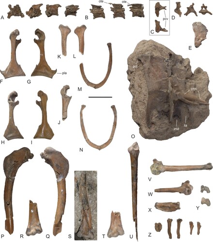
FIGURE 8. Comparison of selected postcranial elements of the Masillaraptoridae, crown group Falconiformes, and crown group Cariamiformes. A–D, coracoid (dorsal view) of A, Masillaraptor cf. parvunguis (NMS.Z.2021.40.14), B, Danielsraptor phorusrhacoides (NMS.Z.2021.40.13), C, Caracara plancus (Falconiformes; SMF 3462), and D, Cariama cristata (Cariamiformes; SMF 1862). E–G, right humerus (caudal view) of E, Ma. cf. parvunguis (NMS.Z.2021.40.14), F, Micrastur ruficollis (Falconiformes; SMF 9812), and G, C. cristata (SMF 1862). H, I, distal end of left humerus (cranial view) of H, Ma. cf. parvunguis (NMS.Z.2021.40.14) and I, Mi. ruficollis (SMF 9812). J–L, proximal end of right ulna (cranial view) of J, Ma. cf. parvunguis (NMS.Z.2021.40.14), K, Mi. ruficollis (SMF 9812), and L, C. cristata (SMF 1862). M–O, left carpometacarpus of M, Ma. cf. parvunguis (NMS.Z.2021.40.15), N, Mi. ruficollis (SMF 9812; right side, mirrored), and O, C. cristata (SMF 1862; right side, mirrored). P–R, pelvis (dorsal view) of P, D. phorusrhacoides (holotype, NMS.Z.2021.40.12; surrounding matrix digitally brightened), Q, Mi. ruficollis (SMF 9812), and R, C. cristata (SMF 1862). S, T, left femur (cranial view) of S, D. phorusrhacoides (holotype, NMS.Z.2021.40.12; surrounding matrix digitally brightened) and T, Mi. ruficollis (SMF 9812). U–AA, proximal (V, X, Z) and distal (U, W, Y, AA) ends of the right tarsometatarsus of U, Ma. cf. parvunguis (NMS.Z.2021.40.14), V, W, D. phorusrhacoides (holotype, NMS.Z.2021.40.12), X, Y, C. plancus (SMF 6441), and Z, AA, C. cristata (SMF 1862). Abbreviations: ctd, cotyla dorsalis, ctr, crista trochanteris; ctv, cotyla ventralis; ecr, insertion scar for musculus extensor carpi radialis; flg, wing-like flange; flx, processus flexorius, fns, foramen nervi supracoracoidei; pex, processus extensorius; pnf, pneumatic foramen; sul, hypotarsal sulcus. Scale bars equal 10 mm.
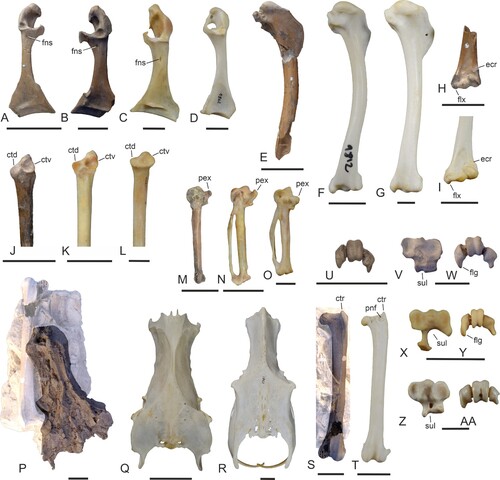
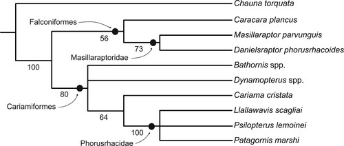
FIGURE 9. Strict consensus tree of the two most parsimonious trees (L = 64, CI = 0.67, RI = 0.73) resulting from the phylogenetic analysis; unsupported nodes were collapsed. Bootstrap support values are indicated next to the internodes.