Figures & data
FIGURE 1. NHMUK PV R37110, the holotype of Ceoptera evansae, approximately as it was found (top) and by CT reconstruction with elements (bottom). Letters indicate the blocks as discussed in the text. Abbreviations: c, coracoid; cv, caudal vertebra; dsc, distal syncarpal; dv, dorsal vertebra; fe, femur; m, manual-phalanx; mc, metacarpal; mt, metatarsal; p, pedal-phalanx; pe, pelvis; ph, phalanx; psc, proximal syncarpal; r, rib; ra, radius; s, scapula; st, sternum; tf, tibia + fibula; ul, ulna; v, vertebra; w, wing-phalanx.
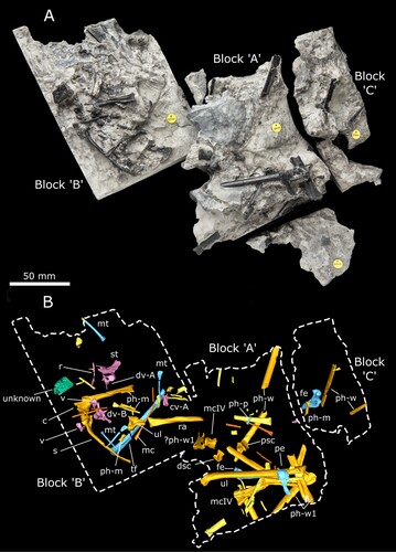
FIGURE 2. Reconstruction of the acetabulum and postacetabular process of the left pelvis of Ceoptera evansae (NHMUK PV R37110), in A, lateral, and B, medial views. Abbreviations: a, acetabulum; dx, dorsal expansion; ic, ischium; pap, postacetabular process; re, recess.
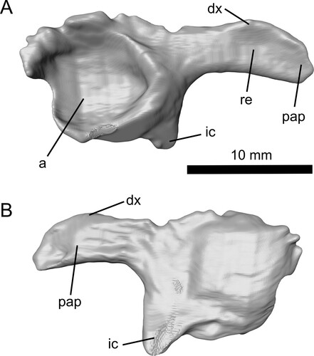
FIGURE 3. The partially preserved right ulna of Ceoptera evansae (NHMUK PV R37110), in A, ventral, and B, dorsal views. Abbreviations: t, tubercle.
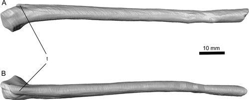
FIGURE 4. Complete left metacarpal IV of Ceoptera evansae (NHMUK PV R37110), in: A, anterior; B, posterior; and C, distal views. Abbreviations: dc, dorsal condyle; vc, ventral condyle; ve, ventral expansion.
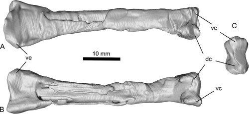
FIGURE 5. Right femur of Ceoptera evansae (NHMUK PV R37110), lacking a short section of the diaphysis, in A, C, anterior, B, D, posterior, and E, distal views. The proximal portion (A, B) is located in Block C while the distal portion (C–E) is located in Block B. Abbreviations: cf, collum femoralis; gr, groove; gt, greater trochanter; mc, medial condyle.
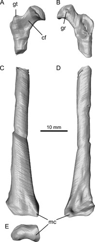
FIGURE 6. Reconstruction of the sternum (primarily the cristospine) of Ceoptera evansae (NHMUK PV R37110), in A, dorsal, B, ventral, and C, left lateral views made from CT scans. Abbreviations: cs, cristospine; f, foramen; lcf, left coracoid facet; rcf, right coracoid facet; sp, sternal plate.
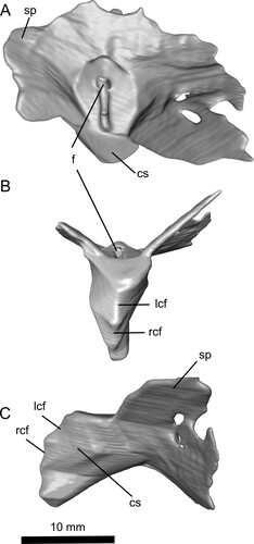
FIGURE 7. Dorsal vertebrae A (A, B) and B (C, D) of Ceoptera evansae (NHMUK PV R37110), in A, C, anterior and B, D, posterior views. Abbreviations: c, centrum; cf, capitular facet; ns, neural spine; prz, prezygopophysis; tf, tubercular facet; tp, transverse process.
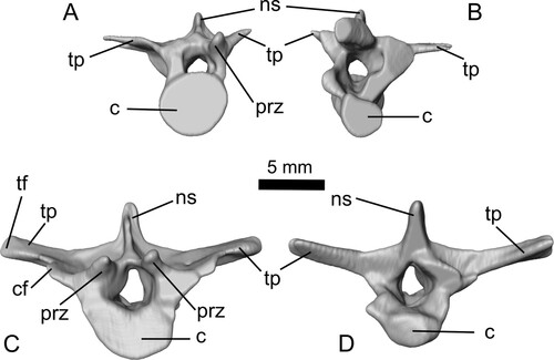
FIGURE 8. Reconstruction of the right scapulocoracoid of Ceoptera evansae (NHMUK PV R37110), made from CT scans in A, lateral and B, medial views. C, reveals a close-up of the expanded sub-triangular brachial flange of the coracoid, one of the diagnostic characters of this taxon. Abbreviations: acc, acrocoracoid process; afs, articular facet for sternum; bf, brachial flange; c, coracoid; cf, coracoid flange; gl, glenoid; s, scapula; sde, distal expansion of scapula. The top scale bar is for A and B.
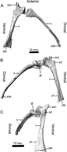
FIGURE 9. Tibia-fibula of Ceoptera evansae (NHMUK PV R37110), in A, lateral, and B, medial views. Abbreviations: fi, fibula; ti, tibia.
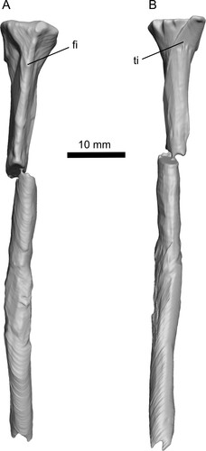
FIGURE 10. Skeletal of Ceoptera evansae (NHMUK PV R37110), showing the material that is present (top, with grayed bones indicating partially preserved elements) and an artist's impression of what the entire skeleton would have looked like if complete. Image copyright Mark Witton.
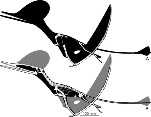
FIGURE 11. Reduced strict consensus tree of 2890 most parsimonious trees (tree lengths = 553; Retention Index = 0.779, Consistency Index = 0.360, with bootstrap/Bremer support values for each node) showing Ceoptera evansae as a basally branching monofenestratan (M) pterosaur within Darwinoptera (D). Silhouettes from top to bottom of: Preondactylus; Jeholopterus; Rhamphorhynchus; Darwinopterus; Ctenochasma; Nyctosaurus; Germanodactylus; and Quetzalcoatlus. Silhouettes from PhyloPic, attributions can be found in the Supplementary Material S1, section 9. See text and Supplementary Material 1, sections 3 and 6–8 for further detail.
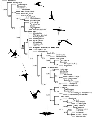
FIGURE 12. Life reconstruction of Ceoptera evansae. Image copyright Mark Witton.