
Figures & data
FIGURE 1. Tooth plate terminology used in this paper. A, orientation of medial, labial, and lingual margins of a tooth plate. B, terminology of parts of a tooth plate. Gray area represents approximate region of occlusal surface. C, lateral view of the end (at the labial edge) of a single tooth ridge showing unworn cusps.
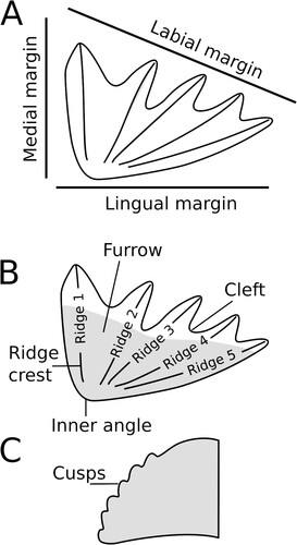
FIGURE 2. Ferganoceratodus edwardsi. A-E, upper tooth plates; F-J, lower tooth plates. A-B, NHMZ 2432(9), holotype in labial and occlusal views. Arrowhead indicates the surface for a potential symphyseal contact; C, NHMZ 2432(6); D, NHMZ 2432(13); E, NHMZ 2432(5); F, NHMZ 2432(1), juvenile; G, NHMZ 2432(2), juvenile; H, NHMZ 2432(12); I, NMHZ 2432(11); J, NHMZ 2432(14); K, NHMZ 2432(4), juvenile. Scale bars equal 10 mm.
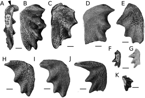
FIGURE 3. Petrological micrographs of Ferganoceratodus edwardsi (NHMZ 2432[8], prearticular tooth plate). A, plane-polarized light (PPL) coronal section through heel of tooth plate showing pulp canals and dentinal tubules below the occlusal surface surrounded by circumdenteonal dentine. Interdenteonal dentine is present between denteons and a thin layer of probable marginal dentine lies just below the occlusal surface; B, section in ‘A’ in crossed-polarized light (XPL) showing inclined orientation of interdenteonal dentine crystals and monorefringent layer of marginal dentine below occlusal surface; C, median section below occlusal surface in PPL through fourth tooth ridge showing inverted ‘cones’ predominantly of circumdenteonal dentine with branching denteons and regions of interdenteonal dentine between cones; D, ‘C’ in XPL; E, ridge of harder interdenteonal dentine with softer ‘cones’ of circumdenteonal dentine on either side. The denteons in the interdenteonal dentine converge towards the occlusal surface; F, ‘D’ in XPL; G, median section through end of tooth ridge showing thin layer of enamel on surface of tooth ridge and complex association of layers of monorefringent and birefringent interdenteonal dentine among a cone of circumdenteonal dentine. Note how denteons are subparallel to the surface of the tooth ridge below the region covered with enamel; H, XPL of ‘G’ overlain with transparency of ‘G’ to highlight relationship between birefringent interdenteonal dentine and regions of darker dentine seen in PPL. Abbreviations: cd, circumdenteonal dentine; e, enamel; id, interdenteonal dentine; md, marginal dentine; t, dentinal tubules; pc, pulp canal. Scale bars for A, B, E, F equal 500 μm. Scale for C, D, G, H equal 1 mm.
![FIGURE 3. Petrological micrographs of Ferganoceratodus edwardsi (NHMZ 2432[8], prearticular tooth plate). A, plane-polarized light (PPL) coronal section through heel of tooth plate showing pulp canals and dentinal tubules below the occlusal surface surrounded by circumdenteonal dentine. Interdenteonal dentine is present between denteons and a thin layer of probable marginal dentine lies just below the occlusal surface; B, section in ‘A’ in crossed-polarized light (XPL) showing inclined orientation of interdenteonal dentine crystals and monorefringent layer of marginal dentine below occlusal surface; C, median section below occlusal surface in PPL through fourth tooth ridge showing inverted ‘cones’ predominantly of circumdenteonal dentine with branching denteons and regions of interdenteonal dentine between cones; D, ‘C’ in XPL; E, ridge of harder interdenteonal dentine with softer ‘cones’ of circumdenteonal dentine on either side. The denteons in the interdenteonal dentine converge towards the occlusal surface; F, ‘D’ in XPL; G, median section through end of tooth ridge showing thin layer of enamel on surface of tooth ridge and complex association of layers of monorefringent and birefringent interdenteonal dentine among a cone of circumdenteonal dentine. Note how denteons are subparallel to the surface of the tooth ridge below the region covered with enamel; H, XPL of ‘G’ overlain with transparency of ‘G’ to highlight relationship between birefringent interdenteonal dentine and regions of darker dentine seen in PPL. Abbreviations: cd, circumdenteonal dentine; e, enamel; id, interdenteonal dentine; md, marginal dentine; t, dentinal tubules; pc, pulp canal. Scale bars for A, B, E, F equal 500 μm. Scale for C, D, G, H equal 1 mm.](/cms/asset/4c1537c2-6b98-4362-a722-3c256fe2336a/ujvp_a_2365391_f0003_oc.jpg)
FIGURE 4. A, vertical micro-CT section though the heel of prearticular tooth plate NHMZ 2432(8). Medial margin is to the left showing high X-ray opacity circumdenteonal dentine and a thin layer of enamel on the surface. ‘Cones’ of higher X-ray opacity with well-developed circumdenteonal dentine coincide with wear pits in the occlusal surface (top) while the main body of the pulp canal is composed of interdenteonal dentine with denteons dispersed throughout. Line represents section in ‘B’; B, horizontal micro-CT section through NHMZ 2432(8) showing regions of pits, and depressions corresponding to circumdenteonal dentine (high X-ray opacity). Abbreviations: cd, circumdenteonal dentine; e, enamel; id, interdenteonal dentine; md, marginal dentine. Scale bars equal 2 mm.
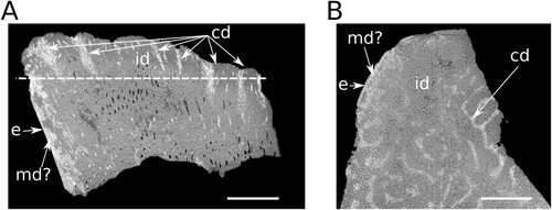
FIGURE 5. Comparison of Ferganoceratodus erdwardsi tooth plates with the type species of Ptychoceratodus, P. serratus. A, holotype of P. serratus (redrawn from Agassiz, Citation1833–43:vol. 3, pl. 19, fig. 18). B, material referred to P. serratus by Schultze (Citation1981). C, the new material of F. edwardsi. D, the type series of Ferganoceratodus, F. jurassicus (redrawn from Nessov & Kaznyshkin, Citation1985). E, type specimen of F. phillipsi upper row, pterygoid tooth plates; lower row, prearticular tooth plates. Scale bars equal 10 mm.
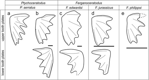
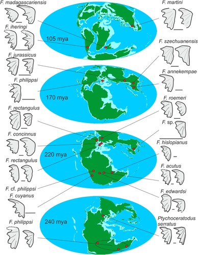
FIGURE 6. Geographic and stratigraphic distributions of Ferganoceratodus spp. and Ptychoceratodus serratus. See text for details.
Supplemental Material
Download MS Word (7.2 MB)DATA AVAILABILITY STATEMENT
The data that support the findings of this study are openly available in Morphosource at https://www.morphosource.org, project ID: 000547763 and https://doi.org/10.17602/M2/M547805
.