Figures & data
FIG. 1 Schematic of an idealized particle with a simple radially symmetrical structure.
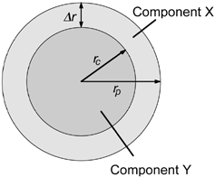
FIG. 2 Schematic of a simple radially symmetrical particle with mixing of components X and Y between the core and shell.
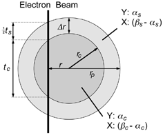
FIG. 3 Schematic of the system used to generate and collect welding fume.
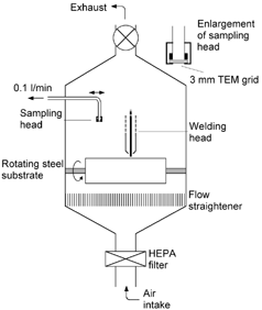
FIG. 4 STEM annular dark field images of collected welding fume, showing the presence of two distinct types of primary particle.
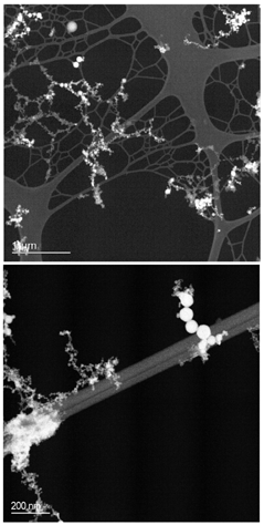
FIG. 5 Conventional TEM images, showing the presence of a thin outer shell-like structure around larger welding fume primary particles. Lattice fringes are apparent in the top image, showing the core of the particle to be crystalline.
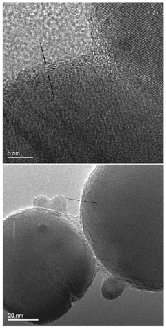
FIG. 6 Energy-filtered image of the edge of two adjoining particles, indicating a region of high Si content (bright feature).
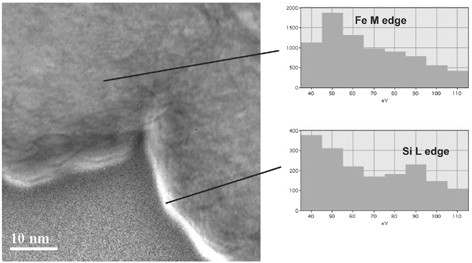
FIG. 7 STEM annular dark field image of a welding particle characterized using EDS. Analysis was carried out at the points shown using a 0.5 nm diameter electron beam and with acquisition times typically of 60 s.
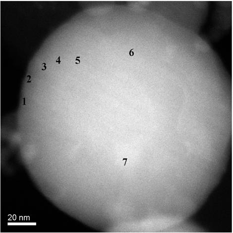
FIG. 8 STEM annular dark field image of welding particle used in EELS analysis. Analysis was carried out in the regions shown using a 0.2 nm diameter electron beam and with acquisition times between of 3 and 6 s.
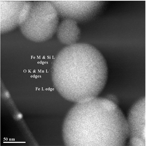
FIG. 9 EELS spectra from the edge of a 5 nm particle. The exponentially decreasing energy-loss background has been removed from both edges shown, allowing qualitative comparison of the Si/Fe ratio with analysis position. Spectra from positions 1–3 are vertically offset to allow easier comparison.
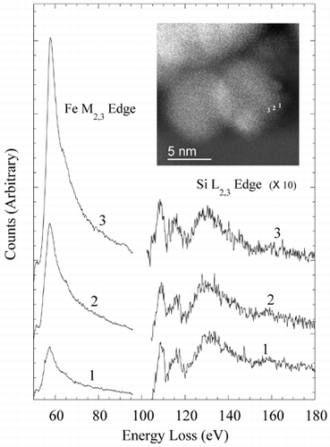
FIG. 10 Elemental ratio (X-ray counts) as a function of radial distance for the particle shown in , measured using EDS analysis. EquationEquation (5) has been fit to the data using nonlinear regression to estimate the various model parameters and shell thickness λ r. In each case λ c and λ s have been assigned nominal values of 1. Estimated parameter errors are the standard error from the nonlinear regression.
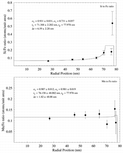
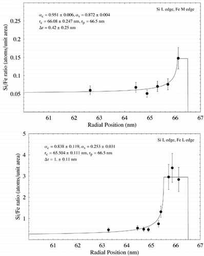
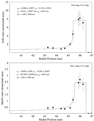
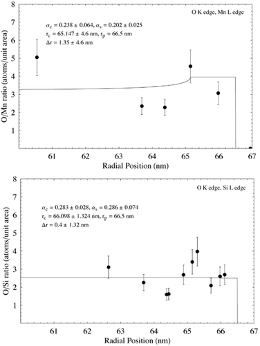
FIG. 11 Elemental ratio (atoms per unit area) as a function of radial distance for the particle shown in measured using EELS analysis. EquationEquation (5) has been fit to the data using nonlinear regression to estimate the various model parameters and shell thickness λ r. In each case λ c and λ s have been assigned nominal values of 1. Estimated parameter errors are associated with uncertainty over radial position, and the model fit. Data points are shown with an estimated ± 20% error. (Continued)