Figures & data
TABLE 1 Summary of experiments performed
FIG. 1a (a) Expanded view of the electrostatic precipitator with mounted X-ray emitter. (Continued)
FIG. 1b (b) Experimental setup used to measure size distribution, charge, and capture efficiency of MS2 Virion particles in a soft X-ray enhanced electrostatic precipitator. FCE, Faraday Cup Electrometer.
FIG. 2 Current-voltage characteristics of the ESP with positive and negative applied potentials.
FIG. 3 MS2 particle size distribution aerosolized by (a) a Collison nebulizer without neutralizer, (b) stainless steel atomizer without neutralizer, (c) nebulizer with neutralizer, (d) nebulizer with soft X-ray, and (e) nebulizer with neutralizer and soft X-ray. The SMPS system had an impactor to remove large agglomerates prior to entry into the Kr-85 neutralizer, and a negative voltage was applied to the central DMA electrode.

TABLE 2 Integral properties of measured size distributions under a variety of conditions (Set 2, )
FIG. 4 SEM images of MS2 agglomerates: (a) suspensions in solution, (b) airborne virion particles after nebulization.
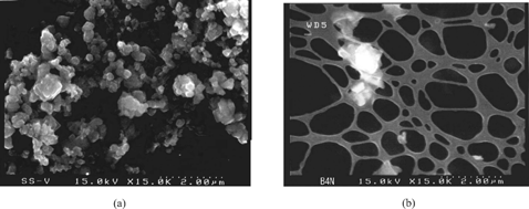
FIG. 5 (a) Electrometer current measured for different particle electrical mobilities set using a DMA. (b) Particle concentrations at different particle electrical mobilities set using a DMA.
FIG. 6 Calculated average particle charge at the ESP outlet with varying applied voltages from −10 to 10 kV.
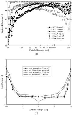
FIG. 7 (a) Capture efficiency of different particle diameter viral particles from 10 to 225 nm for various applied voltages and soft X-ray irradiation. (b) Log capture of viral aerosols as a function of applied voltage.