Figures & data
FIG. 1 Schematic view of the EFR, refractory tube, and chimney.
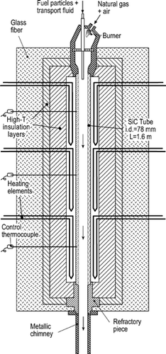
FIG. 2 Measured gas temperature evolution with time along the lower part of the heated tube, refractory section, and chimney. The sampling points used for studying the particle formation process are marked.
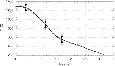
FIG. 3 Schematic view of the AQPS probe.
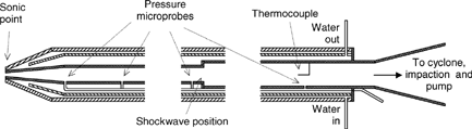
FIG. 4 Measured and predicted pressure and static temperature evolution with length along the AQPS probe.
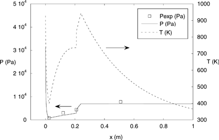
TABLE 1 Proximate and ultimate ASTM analysis and inorganic matter composition of the sieved orujillo
FIG. 5 Particle size distribution of the final emissions. Note the difference between the scales for both modes. (dLog10Dp equals 0.0664 for the laser diffractometer and ∼0.31 for the BLPI).
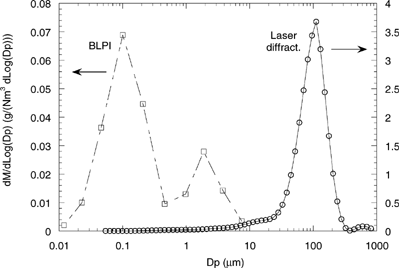
FIG. 6 DMA-derived PSD distribution (mass basis) compared with the BLPI result. Both series have been normalized by their peak height.
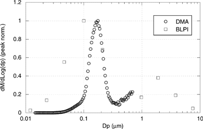
FIG. 7 Chemical composition (in a mass basis) of deposits on BLPI stages 1 through 6, and 8 and 9, compared with the mineral matter composition of the original fuel.
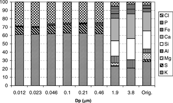
FIG. 8 X-ray diffractogram of a sample from stage 4 of the BLPI (dp = 100 nm). The aluminum substrate peaks are identified.
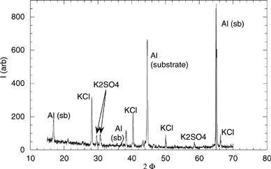
FIG. 9 SEM micrographs of deposits in cyclone (a), and BLPI stages 9 (b), and 4 (c).
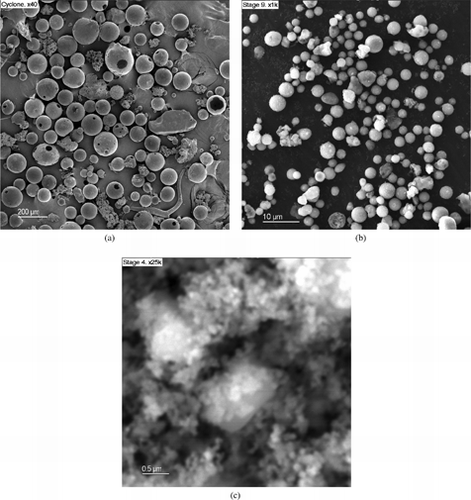
FIG. 10 Comparison between the PSD of ash and original fuel.
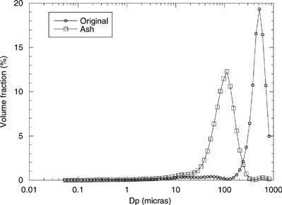
FIG. 11 TEM pictures of samples collected in the EFR using the AQPS probe at (a) 1300°C, (b) 900°C, and (c) 560°C. (Continued)
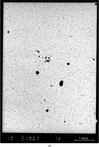
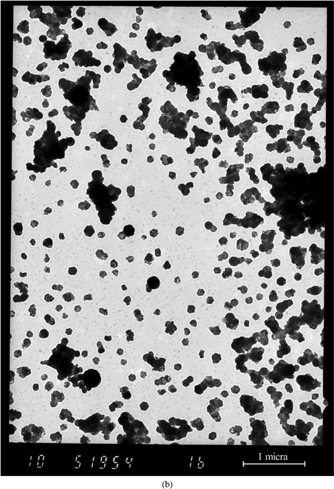
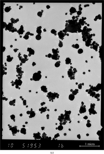
FIG. 12 TEM-XEDS spectra of samples collected at 900°C and 560°C. Both curves have been normalized by their K (Kα) peak height. The small peaks to the left of P are spurious due to the measurement technique.
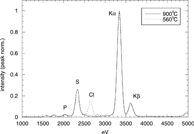
TABLE 2 Input data and extract of the species considered in the thermodynamic equilibrium calculations
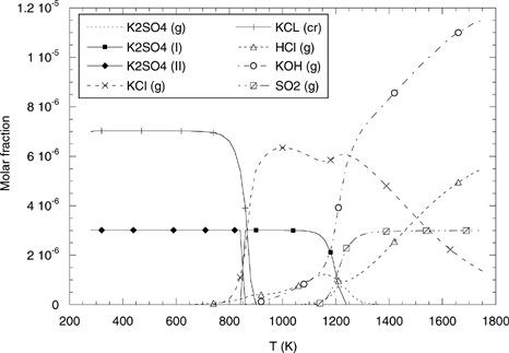
FIG. 13 Equilibrium distribution of gaseous (dashed curves) and condensed (continuous curves) species in the EFR as a function of temperature.